
Page 1667 - Williams Hematology ( PDFDrive )
P. 1667
1642 Part XI: Malignant Lymphoid Diseases Chapter 99: Follicular Lymphoma 1643
A B
C D E
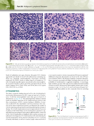
Figure 99–1. Follicular lymphoma grading is based on the relative proportions of small cells (centrocytes) and centroblasts (centroblasts). A. Grade 1
(0–5 centroblasts/high-powered field). B. Grade 2 (6–15 centroblasts/high-powered field). C. Grade 3A (>15 centroblasts/high-powered field).
D and E. Grade 3B. See text for further definitions of grades 1, 2, 3A, and 3B. (Reproduced with permission from Harris NL, Swerdlow SH, Jaffe ES, et al:
Follicular Lymphoma, in WHO Classification of Tumours of Haematopoietic and Lymphoid Tissues, edited by Swerdlow SH, Campo E, Harris NL, et al:
p 220–226. International Agency for Research on Cancer, Lyon, 2008.)
Nearly all authorities now agree, however, that grade 3B FL behaves a very sensitive nested or reverse-transcription PCR assay is employed.
2
aggressively and should be treated with anthracycline-containing reg- Additional cytogenetic abnormalities are found in the cells of 90 per-
imens (e.g., rituximab, cyclophosphamide, doxorubicin, vincristine, cent of patients with FL. The finding of multiple cytogenetic abnormal-
prednisone [R-CHOP]) similar to diffuse large B-cell lymphoma. FL ities is commonly associated with higher histologic grade and with the
2
cells of all grades typically express monoclonal surface immunoglobu- probability of transformation to aggressive lymphoma. A recent large,
lin, are positive for BCL-2, BCL6, and CD10, and express the pan–B-cell high-resolution, genome-wide copy-number analysis demonstrated that
surface antigens CD19, CD20, CD22, and CD79a, but do not express common recurrent chromosomal abnormalities include gains of chro-
CD5, CD23, CD11c, or CD43. mosomes 2, 5, 6p, 7, 8, 12, 17q, 18, 21, and X and losses on 6q and 17p.
10
Frequent small abnormalities are also commonly observed, including
CYTOGENETICS
The classic cytogenetic finding detected in FL is the t(14;18)(q32;q21)
translocation that juxtaposes the BCL-2 gene on band q21 of chromo- 18q –
some 18 with the immunoglobulin heavy-chain gene on band 32 of p p
9
chromosome 14 (Fig. 99–2). The immunoglobulin enhancer element
results in amplified expression of the translocated gene product and,
thus, overexpression of BCL-2 protein leading to inhibition of apop-
tosis of affected B cells. Quantitative real-time polymerase chain reac- BCL-2
tion (PCR) assays on blood and marrow can determine the number of q q H-chain
t(14;18)-expressing cells and may be useful in predicting the outcome of # 18 enhancer
therapy. The t(14;18) translocation is found in approximately 85 percent Antibody H BCL-2
chains
of patients in the United States, but the translocation is present in a sig-
nificantly lower percentage of Asian patients afflicted with FL. Detection 14q +
of the t(14;18) translocation in lymphoid cells is neither necessary nor # 14
sufficient for the diagnosis of FL. Small numbers of B cells harboring the Figure 99–2. The t(14;18)(q32;q21) translocation juxtaposes the BCL-2
t(14;18) translocation can be detected in the blood of 25 to 75 percent gene on band q21 of chromosome 18 with the immunoglobulin heavy-
of healthy individuals, as well as in reactive lymph nodes and tonsils if chain gene on band 32 of chromosome 14.
Kaushansky_chapter 99_p1641-1652.indd 1642 9/18/15 3:57 PM