
Page 1698 - Williams Hematology ( PDFDrive )
P. 1698
1672 Part XI: Malignant Lymphoid Diseases Chapter 102: Burkitt Lymphoma 1673
TABLE 102–1. Murphy Staging System for Burkitt LABORATORY FEATURES
Lymphoma
Stage I: Single nodal or extranodal site excluding mediastinum or BLOOD AND MARROW
abdomen Patients with bulky disease may have Burkitt cells in marrow and blood
Stage II: Single extranodal tumor with regional nodal involvement with accompanying suppression of normal blood counts. Characteristic
pathologic features of BL on smear preparation are intermediate-size cells
Two extranodal tumors on one side of diaphragm with round nuclei, multiple nucleoli, strongly basophilic cytoplasm (a
Primary gastrointestinal tumor with or without associated consequence of the abundant polyribosomes), and the presence of lipid-
mesenteric nodes filled cytoplasmic vesicles, some of which overlie the nucleus (Fig. 102–1).
Two or more nodal areas on one side of diaphragm Rare cases, more commonly in males, may present principally with mar-
Stage IIR: Completely resected intraabdominal disease row and blood involvement, so-called Burkitt cell leukemia variant (pre-
Stage III: Two single extranodal tumors on opposite sides of diaphragm viously classified as acute lymphocytic leukemia–L3, according to the
All primary intrathoracic tumors former French-American-British [FAB] classification).
The serum lactic dehydrogenase (LDH) is often elevated as a
All paraspinal or epidural tumors reflection of the high cell turnover, especially in patients with bulky
All extensive primary intraabdominal disease disease.
Two or more nodal areas on opposite sides of diaphragm
Stage IIIA: Localized, nonresectable abdominal disease HISTOPATHOLOGY AND CYTOLOGY
Stage IIIB: Widespread multiorgan abdominal disease BL is characterized by monomorphic medium-size cells with round
Stage IV: Initial CNS or marrow involvement (<25%) nuclei, multiple nucleoli, and basophilic cytoplasm. Burkitt cells
37
Adapted with permisison from Perkins AS, Friedberg JW: Burkitt have a very high proliferative rate (≥95 percent as determined by Ki-67
lymphoma in adults. Hematology Am Soc Hematol Educ Program staining) and frequent mitotic figures are usually present. BL has a dif-
341–348, 2008. fuse pattern of growth comprised of intermediate-size B cells (12 μM
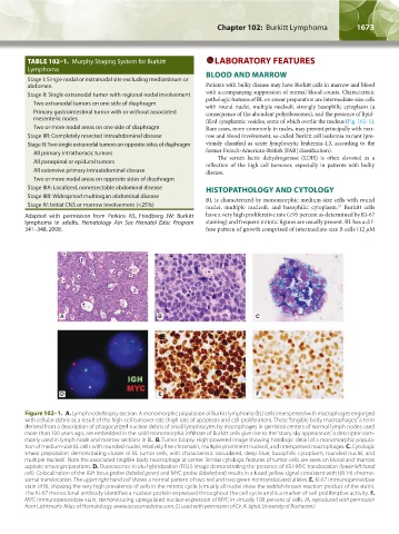
A B C
D E F
Figure 102–1. A. Lymph node biopsy section. A monomorphic population of Burkitt lymphoma (BL) cells interspersed with macrophages engorged
with cellular debris as a result of the high cell turnover rate (high rate of apoptosis and cell proliferation). These “tingible body macrophages,” a term
derived from a description of phagocytized nuclear debris of small lymphocytes by macrophages in germinal centers of normal lymph nodes used
more than 100 years ago, are embedded in the solid monomorphic infiltrate of Burkitt cells give rise to the “starry sky appearance,” a descriptor com-
monly used in lymph node and marrow sections in BL. B. Tumor biopsy. High-powered image showing histologic detail of a monomorphic popula-
tion of medium-size BL cells with rounded nuclei, relatively fine chromatin, multiple prominent nucleoli, and interspersed macrophages. C. Cytologic
smear preparation demonstrating cluster of BL tumor cells, with characteristic vacuolated, deep-blue, basophilic cytoplasm, rounded nuclei, and
multiple nucleoli. Note the associated tingible body macrophage at center. Similar cytologic features of tumor cells are seen on blood and marrow
aspirate smear preparations. D. Fluorescence in situ hybridization (FISH) image demonstrating the presence of IGH-MYC translocation (lower left hand
cell). Colocalization of the IGH locus probe (labeled green) and MYC probe (labeled red) results in a fused yellow signal consistent with t(8:14) chromo-
somal translocation. The upper right hand cell shows a normal pattern of two red and two green nontranslocated alleles. E. Ki-67 immunoperoxidase
stain of BL showing the very high prevalence of cells in the mitotic cycle (virtually all nuclei show the reddish-brown reaction product of the stain).
The Ki-67 monoclonal antibody identifies a nuclear protein expressed throughout the cell cycle and is a marker of cell proliferative activity. F.
MYC immunoperoxidase stain, demonstrating upregulated nuclear expression of MYC in virtually 100 percent of cells. (A, reproduced with permission
from Lichtman’s Atlas of Hematology, www.accessmedicine.com. D, used with permission of Dr. A. Iqbal, University of Rochester.)
Kaushansky_chapter 102_p1671-1678.indd 1673 17/09/15 3:20 pm