
Page 1829 - Williams Hematology ( PDFDrive )
P. 1829
1804 Part XI: Malignant Lymphoid Diseases Chapter 110: Heavy-Chain Disease 1805
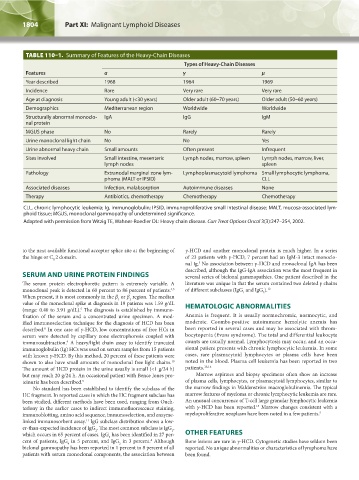
TABLE 110–1. Summary of Features of the Heavy-Chain Diseases
Types of Heavy-Chain Diseases
Features α γ μ
Year described 1968 1964 1969
Incidence Rare Very rare Very rare
Age at diagnosis Young adult (<30 years) Older adult (60–70 years) Older adult (50–60 years)
Demographics Mediterranean region Worldwide Worldwide
Structurally abnormal monoclo- IgA IgG IgM
nal protein
MGUS phase No Rarely Rarely
Urine monoclonal light chain No No Yes
Urine abnormal heavy chain Small amounts Often present Infrequent
Sites involved Small intestine, mesenteric Lymph nodes, marrow, spleen Lymph nodes, marrow, liver,
lymph nodes spleen
Pathology Extranodal marginal zone lym- Lymphoplasmacytoid lymphoma Small lymphocytic lymphoma,
phoma (MALT or IPSID) CLL
Associated diseases Infection, malabsorption Autoimmune diseases None
Therapy Antibiotics, chemotherapy Chemotherapy Chemotherapy
CLL, chronic lymphocytic leukemia; Ig, immunoglobulin; IPSID, immunoproliferative small intestinal disease; MALT, mucosa-associated lym-
phoid tissue; MGUS, monoclonal gammopathy of undetermined significance.
Adapted with permission from Witzig TE, Wahner-Roedler DL: Heavy chain disease. Curr Treat Options Oncol 3(3):247–254, 2002.
to the next available functional acceptor splice site at the beginning of γ-HCD and another monoclonal protein is much higher. In a series
the hinge or C 2 domain. of 23 patients with γ-HCD, 7 percent had an IgM-λ intact monoclo-
H
5
nal Ig. No association between γ-HCD and monoclonal IgA has been
described, although the IgG-IgA association was the most frequent in
SERUM AND URINE PROTEIN FINDINGS several series of biclonal gammopathies. One patient described in the
The serum protein electrophoretic pattern is extremely variable. A literature was unique in that the serum contained two deleted γ chains
monoclonal peak is detected in 60 percent to 86 percent of patients. of different subclasses (IgG and IgG ). 12
4,5
2
1
When present, it is most commonly in the β or β region. The median
2
1
value of the monoclonal spike at diagnosis in 19 patients was 1.59 g/dL HEMATOLOGIC ABNORMALITIES
(range: 0.40 to 3.91 g/dL). The diagnosis is established by immuno-
5
fixation of the serum and a concentrated urine specimen. A mod- Anemia is frequent. It is usually normochromic, normocytic, and
ified immunoselection technique for the diagnosis of HCD has been moderate. Coombs-positive autoimmune hemolytic anemia has
described. In one case of γ-HCD, low concentrations of free HCs in been reported in several cases and may be associated with throm-
8
serum were detected by capillary zone electrophoresis coupled with bocytopenia (Evans syndrome). The total and differential leukocyte
immunosubtraction. A heavy/light chain assay to identify truncated counts are usually normal. Lymphocytosis may occur, and an occa-
9
immunoglobulin (Ig) HCs was used on serum samples from 15 patients sional patient presents with chronic lymphocytic leukemia. In some
with known γ-HCD. By this method, 20 percent of these patients were cases, rare plasmacytoid lymphocytes or plasma cells have been
shown to also have small amounts of monoclonal free light chains. noted in the blood. Plasma cell leukemia has been reported in two
10
The amount of HCD protein in the urine usually is small (<1 g/24 h) patients. 13,14
but may reach 20 g/24 h. An occasional patient with Bence Jones pro- Marrow aspirates and biopsy specimens often show an increase
teinuria has been described. 5 of plasma cells, lymphocytes, or plasmacytoid lymphocytes, similar to
No standard has been established to identify the subclass of the the marrow findings in Waldenström macroglobulinemia. The typical
HC fragment. In reported cases in which the HC fragment subclass has marrow features of myeloma or chronic lymphocytic leukemia are rare.
been studied, different methods have been used, ranging from Ouch- An unusual concurrence of T-cell large granular lymphocytic leukemia
15
terlony in the earlier cases to indirect immunofluorescence staining, with γ-HCD has been reported. Marrow changes consistent with a
immunoblotting, amino acid sequence, immunoselection, and enzyme- myeloproliferative neoplasm have been noted in a few patients. 5
linked immunosorbent assay. IgG subclass distribution shows a low-
11
er-than-expected incidence of IgG . The most common subclass is IgG ,
2
1
which occurs in 65 percent of cases. IgG has been identified in 27 per- OTHER FEATURES
3
cent of patients, IgG in 5 percent, and IgG in 3 percent. Although Bone lesions are rare in γ-HCD. Cytogenetic studies have seldom been
4
4
2
biclonal gammopathy has been reported in 1 percent to 8 percent of all reported. No unique abnormalities or characteristics of lymphoma have
patients with serum monoclonal components, the association between been found.
Kaushansky_chapter 110_p1803-1812.indd 1804 9/18/15 9:57 AM