
Page 1832 - Williams Hematology ( PDFDrive )
P. 1832
1806 Part XI: Malignant Lymphoid Diseases Chapter 110: Heavy-Chain Disease 1807
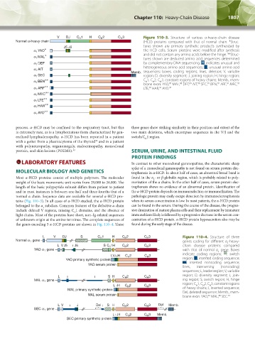
Figure 110–3. Structure of various α-heavy-chain disease
- (HCD) proteins compared with that of normal chain. *Struc-
tures shown are primary synthetic products synthesized by
the HCD cells. Serum proteins were modified after synthesis
and did not contain any amino acids before the hinge. **Struc-
tures shown are deduced amino acid sequences determined
by complementary DNA sequencing. H, indicates unusual and
heterogeneous amino acid sequences; , unusual amino acid
sequences; boxes, coding regions; lines, deletion; V, variable
region; D, diversity segment; J, joining region; H, hinge region;
C 1, C 2, C 3, constant regions of heavy chains; Memb., mem-
H
H
H
92
91
92
92
90
87
brane exon. YAO, MAL, DEF, AIT, SEC, BEN, ARF, MEC,
89
88
LTE, HAR, AYO. 92
92
92
process. α-HCD may be confined to the respiratory tract, but this three genes show striking similarity in their position and extent of the
is extremely rare, as is a lymphomatous form characterized by gen- two main deletions, which encompass sequences in the V/J and the
eralized lymphadenopathy. α-HCD has been reported in a patient switch/C 1 region.
H
with a goiter from a plasmacytoma of the thyroid and in a patient
32
with polyneuropathy, organomegaly, endocrinopathy, monoclonal
protein, and skin lesions (POEMS). 33 SERUM, URINE, AND INTESTINAL FLUID
PROTEIN FINDINGS
LABORATORY FEATURES In contrast to other monoclonal gammopathies, the characteristic sharp
spike of a monoclonal gammopathy is not found on serum protein elec-
MOLECULAR BIOLOGY AND GENETICS trophoresis in α-HCD. In about half of cases, an abnormal broad band is
Most α-HCD proteins consist of multiple polymers. The molecular found in the α - or β-globulin region, which is probably related to poly-
2
weight of the basic monomeric unit varies from 29,000 to 34,000. The merization of the α chains. In the other half of cases, serum protein elec-
length of the basic polypeptide subunit differs from patient to patient trophoresis shows no evidence of an abnormal protein. Identification of
and in most instances is between one-half and three-fourths that of a the α-HCD protein depends on immunoselection or immunofixation. The
normal α chain. Sequence data are available for several α-HCD pro- pathologic protein may easily escape detection by immunoelectrophoresis
teins (Fig. 110–3). In all cases of α-HCD studied, the α-HCD protein when its serum concentration is low. In most patients, the α-HCD protein
belonged to the α subclass. Common features of the defective α chain can be found in the serum. During the course of the disease, the progres-
1
include deleted V regions, missing C 1 domains, and the absence of sive diminution of mature plasma cells and their replacement by immature
H
light chains. Most of the proteins have short, non–Ig-related sequences immunoblasts likely is followed by a progressive decrease in the serum con-
of unknown origin at the amino terminus. The complete sequences of centration of α-HCD protein. α-HCD protein hyposecretion also may be
the genes encoding 3 α-HCD proteins are shown in Fig. 110–4. These found during the early stage of the disease.
Figure 110–4. Structure of three
genes coding for different α -heavy-
1
chain disease proteins compared
with that of normal α gene. Boxes
1
indicate coding regions; , switch
region; , inserted coding sequence;
, inserted noncoding sequence;
lines, intervening (noncoding)
sequences; L, leader region; V, variable
region; D, diversity segment; J, join-
ing region; S, switch region; H, hinge
region; C 1, C 2, C 3, constant regions
H
H
H
of heavy chains; I, inserted sequence;
Del, deleted sequence; Memb., mem-
88
87
brane exon. YAO, MAL, SEC. 91
Kaushansky_chapter 110_p1803-1812.indd 1807 9/18/15 9:58 AM