
Page 1841 - Williams Hematology ( PDFDrive )
P. 1841
1816 Part XII: Hemostasis and Thrombosis Chapter 111: Megakaryopoiesis and Thrombopoiesis 1817
TABLE 111–1. Maturation Stages of Megakaryocytes not appear to be required for the GPIb-V-IX complex to function as a
von Willebrand factor receptor. Rather, GPV is a target of thrombin,
Term Size (μM) Morphology potentially playing a role in platelet activation. 15
Megakaryoblast >10 Lobed nucleus, basophilic
(stage I) cytoplasm Demarcation Membranes
Basophilic >20 Horseshoe-shaped nucleus, Another feature of the megakaryoblast is the initial development of
megakaryocyte basophilic cytoplasm, demarcation membranes, which begin as invaginations of the plasma
(stage II) azurophilic granules around membrane and ultimately develop into a highly branched intercon-
centrosome nected system of channels that course through the cytoplasm. The
demarcation membrane system is in open communication with the
Granular megakary- >25–50 Large multilobed nucleus, aci- 16
ocyte (stage III) dophilic cytoplasm, numer- extracellular space, based on studies using electron dense tracers. Bio-
ous azurophilic granules chemical analysis indicates the composition of these membranes is very
similar to the plasma membrane at each stage of megakaryocyte devel-
Mature megakaryo- >25–50 Pyknotic nucleus, groups of opment. Over the 72 hours required for stage III/IV cells to develop
cyte (stage IV) 10–12 azurophilic granules
from megakaryoblasts, the demarcation membrane system grows sub-
stantially. The demarcation membrane system provides the material
necessary for development of proplatelet processes, structures that form
second most abundant megakaryocyte-specific protein. Glycoprotein V in stage IV megakaryocytes and give rise upon fragmentation to mature
12
also is expressed in complex with GPIb and GPIX, in a ratio of 1:2:2. platelets. 8,17
However, the genetic elimination of GPV has little effect on platelet
13
adhesion, and unlike GPIb and GPIX, no mutations of GPV are asso- Endomitosis
ciated with Bernard-Soulier disease (Chap. 120). Therefore, GPV does One of the most characteristic features of megakaryocyte development
14
is endomitosis, a unique form of mitosis in which the DNA is repeat-
edly replicated in the absence of nuclear or cytoplasmic division. The
resultant cells are highly polyploid. Endomitosis begins in megakary-
oblasts (Fig. 111–2) following the many standard cell divisions required
Pluripotential
stem cell
Meg-CFC
2N 4N 8N 16N 32N 64N
Immature
megakaryocytes
Mature
megakaryocytes
Platelets
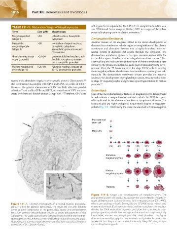
Figure 111–2. Origin and development of megakaryocytes. The
pluripotential stem cell produces a progenitor committed to megakary-
ocyte differentiation (colony-forming unit–megakaryocyte [CFU-MK]),
Figure 111–1. Electron micrograph of a normal human megakary- which can undergo mitosis. Eventually the CFU-MK stops mitosis and
oblast stained for platelet peroxidase. The small cell (<9 μm) exhibits enters endomitosis. During endomitosis, neither cytoplasm nor nucleus
dense platelet peroxidase in the perinuclear space and endoplasmic divides, but DNA replication proceeds and gives rise to immature poly-
reticulum (arrows) (magnification ×12,150). (Inset) Enlargement of the ploid progenitors, which then enlarge and mature into morphologically
Golgi zone. The Golgi saccules and vesicles are devoid of platelet perox- identifiable, mature megakaryocytes that shed platelets. This figure
idase (open arrows), whereas the endoplasmic reticulum contains plate- does not necessarily imply that endomitosis and platelet formation are
let peroxidase activity (closed arrow) (magnification ×25,000). (Used with sequential but they can occur simultaneously. Meg-CFC, megakaryo-
permission of Dr. J. Breton-Gorius.) cyte colony-forming cells.
Kaushansky_chapter 111_p1813-1828.indd 1816 9/21/15 4:11 PM