
Page 1845 - Williams Hematology ( PDFDrive )
P. 1845
1820 Part XII: Hemostasis and Thrombosis Chapter 111: Megakaryopoiesis and Thrombopoiesis 1821
A B C
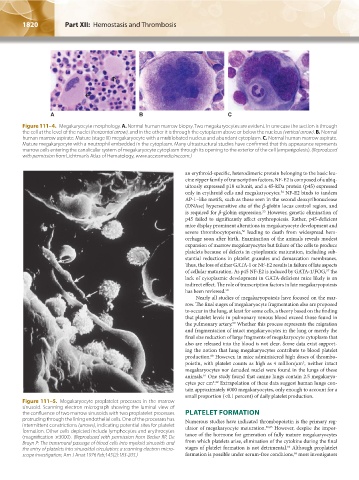
Figure 111–4. Megakaryocyte morphology. A. Normal human marrow biopsy. Two megakaryocytes are evident. In one case the section is through
the cell at the level of the nuclei (horizontal arrow), and in the other it is through the cytoplasm above or below the nucleus (vertical arrow). B. Normal
human marrow aspirate. Mature (stage III) megakaryocyte with a multilobated nucleus and abundant cytoplasm. C. Normal human marrow aspirate.
Mature megakaryocyte with a neutrophil embedded in the cytoplasm. Many ultrastructural studies have confirmed that this appearance represents
marrow cells entering the canalicular system of megakaryocyte cytoplasm through its opening to the exterior of the cell (emperipolesis). (Reproduced
with permission from Lichtman’s Atlas of Hematology, www.accessmedicine.com.)
an erythroid-specific, heterodimeric protein belonging to the basic leu-
cine zipper family of transcription factors, NF-E2 is composed of a ubiq-
uitously expressed p18 subunit, and a 45-kDa protein (p45) expressed
only in erythroid cells and megakaryocytes. NF-E2 binds to tandem
54
AP-1–like motifs, such as those seen in the second deoxyribonuclease
(DNAse) hypersensitive site of the β-globin locus control region, and
is required for β-globin expression. However, genetic elimination of
55
p45 failed to significantly affect erythropoiesis. Rather, p45-deficient
mice display prominent alterations in megakaryocyte development and
severe thrombocytopenia, leading to death from widespread hem-
56
orrhage soon after birth. Examination of the animals reveals modest
expansion of marrow megakaryocytes but failure of the cells to produce
platelets because of defects in cytoplasmic maturation, including sub-
stantial reductions in platelet granules and demarcation membranes.
Thus, the loss of either GATA-1 or NF-E2 results in failure of late aspects
of cellular maturation. As p45 NF-E2 is induced by GATA-1/FOG, the
57
lack of cytoplasmic development in GATA-deficient mice likely is an
indirect effect. The role of transcription factors in late megakaryopoiesis
has been reviewed. 58
Nearly all studies of megakaryopoiesis have focused on the mar-
row. The final stages of megakaryocyte fragmentation also are proposed
to occur in the lung, at least for some cells, a theory based on the finding
that platelet levels in pulmonary venous blood exceed those found in
the pulmonary artery. Whether this process represents the migration
59
and fragmentation of intact megakaryocytes in the lung or merely the
final size reduction of large fragments of megakaryocyte cytoplasm that
also are released into the blood is not clear. Some data exist support-
ing the notion that lung megakaryocytes contribute to blood platelet
production. However, in mice administered high doses of thrombo-
60
poietin, with platelet counts as high as 4 million/μm , neither intact
3
megakaryocytes nor denuded nuclei were found in the lungs of these
animals. One study found that canine lungs contain 2.5 megakaryo-
61
cytes per cm . Extrapolation of these data suggest human lungs con-
2 62
tain approximately 6000 megakaryocytes, only enough to account for a
small proportion (<0.1 percent) of daily platelet production.
Figure 111–5. Megakaryocyte proplatelet processes in the marrow
sinusoid. Scanning electron micrograph showing the luminal view of
the confluence of two marrow sinusoids with two proplatelet processes PLATELET FORMATION
protruding through the lining endothelial cells. One of the processes has Numerous studies have indicated thrombopoietin is the primary reg-
intermittent constrictions (arrows), indicating potential sites for platelet ulator of megakaryocyte maturation. 36,63 However, despite the impor-
formation. Other cells depicted include lymphocytes and erythrocytes tance of the hormone for generation of fully mature megakaryocytes
(magnification ×3000). (Reproduced with permission from Becker RP, De from which platelets arise, elimination of the cytokine during the final
Bruyn P: The transmural passage of blood cells into myeloid sinusoids and 64
the entry of platelets into sinusoidal circulation; a scanning electron micro- stages of platelet formation is not detrimental. Although proplatelet
65
scope investigation, Am J Anat 1976 Feb;145(2):183-205.) formation is possible under serum-free conditions, most investigators
Kaushansky_chapter 111_p1813-1828.indd 1820 9/21/15 4:11 PM