
Page 1976 - Williams Hematology ( PDFDrive )
P. 1976
1950 Part XII: Hemostasis and Thrombosis Chapter 114: Control of Coagulation Reactions 1951
imparts special threshold properties to the blood coagulation pathways, the molecular weight, normal plasma concentration, chromosomal
making the coagulant response nonlinearly responsive to stimuli. The- location, and gene structures of these factors are given in Table 114–1.
oretical analysis of blood coagulation as a threshold system suggests Factors Va and VIIIa, as substrates of APC, are also participants in the
there can be an all-or-none response to various levels of stimulation, reactions of the anticoagulant protein C pathway. Moreover, factor V,
depending on the ensemble of activating and inhibitory reactions that but not factor V Leiden, appears to act as an APC cofactor for the inac-
defines upregulation and downregulation of thrombin generation. 21,22 tivation of factor VIIIa (see “Factor V as Activated Protein C Cofactor”
The coagulation system is active, but idling, and is poised for extensive below). 23
and explosive generation of thrombin. Because of synergy among var-
ious cellular and humoral anticoagulant mechanisms that establish a PROTEIN C
threshold system, the presence of multiple coagulation inhibitors with
complementary modes of action prevents massive thrombin generation In 1976, Stenflo designated a bovine plasma vitamin K–dependent pro-
in the absence of a substantial procoagulant stimulus. tein that eluted in the third peak (peak C) from an anion exchange col-
umn as bovine “protein C.” Protein C, actually previously described
24
as the anticoagulant factor autoprothrombin II-A, is a plasma serine
25
HEREDITARY DEFICIENCIES protease zymogen that can be converted to an active serine protease by
ASSOCIATED WITH THROMBOTIC the action of thrombin.
Protein C is synthesized in the liver as a polypeptide precursor of
DISEASE 461 residues, with a prepropeptide of 42 amino acids that contains the
signal for carboxylation of Glu residues by a carboxylase that forms nine
Evidence for the physiologic importance of specific factors for control- γ-carboxyglutamic acid (GLA) residues and secretion of the mature
ling coagulation reactions comes from clinical observations and animal protein. 26–28 The mature glycoprotein of Mr 62,000 contains 419 residues
model studies. Major identified genetic risk factors for venous throm- (see Chap. 113, Fig. 113–1 and Fig. 114–5) and N-linked carbohydrate,
bosis involve protein structural defects in factor V, protein C, protein and the majority of the secreted protein C molecules are cleaved by a
S, and antithrombin (Chap. 130). There are also gene regulatory defects furin-like endoprotease that releases Lys156-Arg157 and generates a
associated with thrombotic disease, such as the G20210A polymorphism two-chain zymogen that circulates in plasma at 65 nM (4 mcg/mL).
29
in the prothrombin gene that causes elevated levels of prothrombin, and The heavy and light chains of plasma protein C are covalently linked by
defects in protein C gene regulatory elements that decrease the expres- a disulfide bond that keeps the serine protease globular domain (resi-
sion of protein C. Deficiencies of thrombomodulin might also be associ- dues 170 to 419) covalently tethered to the N-terminal string of three
ated with increased risk of arterial thrombosis. Association of hereditary domains, the GLA domain and the epidermal growth factor (EGF)-like
abnormalities of EPCR with increased risks of thrombosis has been sug- domains EGF1 and EGF2. 26–30
gested, but this remains somewhat controversial. The GLA domain of protein C (residues 1 to 42) and APC is impor-
tant for a number of functions, including binding to phospholipid-
PROTEIN C PATHWAY COMPONENTS containing membranes (see Chap. 113, Fig. 113–3), thrombomodulin,
and EPCR; thus, incomplete carboxylation impairs the functional anti-
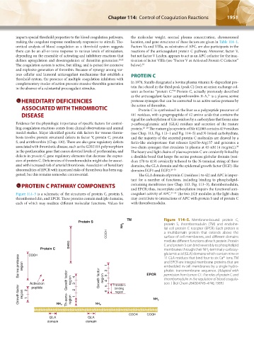
Figure 114–5 is a schematic of the structures of protein C, protein S, coagulant activity of APC. 31–33 The two EGF modules in the light chain
thrombomodulin, and EPCR. These proteins contain multiple domains, may contribute to interactions of APC with protein S and of protein C
each of which may mediate different molecular functions. Values for with thrombomodulin.
TM Figure 114–5. Membrane-bound protein C,
Protein S protein S, thrombomodulin (TM) and endothe-
NH 2 lial cell protein C receptor (EPCR). Each protein is
COOH a multidomain protein that extends above the
surface of cell membranes, and different domains
SHBG mediate different functions of each protein. Protein
C and protein S can bind reversibly to phospholipid
Protein C membranes through their NH -terminal γ-carboxy-
2
glutamic acid (GLA) domains which contain nine or
COOH
Serine protease region 1 2 EPCR and EPCR are integral membrane proteins that are
11 GLA residues that bind four to six Ca ions. TM
2+
embedded in cell membranes by a single hydro-
phobic transmembrane sequence. (Adapted with
permission from Esmon CT: The roles of protein C and
tion. J Biol Chem 264(9):4743–4746, 1989.)
Activation Growth factor region 4 3 Growth factor region 43 5 Thrombin- thrombomodulin in the regulation of blood coagula-
Growth factor region 2 1 NH Th-sens- region 2 1 NH 6 region NH 2
peptide
binding
2
2
COOH COOH
GLA GLA
domain domain
Kaushansky_chapter 114_p1949-1966.indd 1951 9/18/15 10:05 AM