
Page 2159 - Williams Hematology ( PDFDrive )
P. 2159
2134 Part XII: Hemostasis and Thrombosis Chapter 124: Inherited Deficiencies of Coagulation Factors II, V, V+VIII, VII, X, XI, and XIII 2135
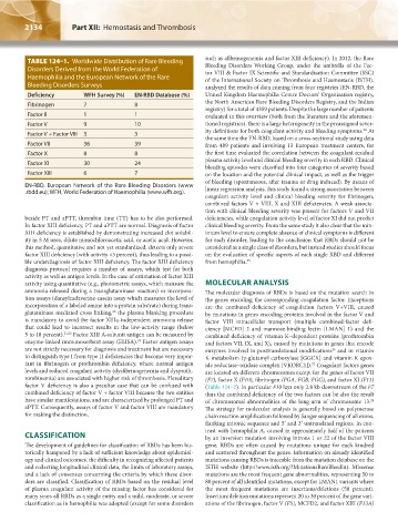
TABLE 124–1. Worldwide Distribution of Rare Bleeding such as afibrinogenemia and factor XIII deficiency). In 2012, the Rare
Bleeding Disorders Working Group, under the umbrella of the Fac-
Disorders Derived from the World Federation of tor VIII & Factor IX Scientific and Standardisation Committee (SSC)
Haemophilia and the European Network of the Rare of the International Society on Thrombosis and Haemostasis (ISTH),
Bleeding Disorders Surveys analyzed the results of data coming from four registries (EN-RBD, the
Deficiency WFH Survey (%) EN-RBD Database (%) United Kingdom Haemophilia Centre Doctors’ Organization registry,
Fibrinogen 7 8 the North American Rare Bleeding Disorders Registry, and the Indian
registry) for a total of 4359 patients. Despite the large number of patients
Factor II 1 1 evaluated in this overview (both from the literature and the aforemen-
Factor V 9 10 tioned registries), there is a large heterogeneity in the preassigned sever-
ity definitions for both coagulant activity and bleeding symptoms. At
24
Factor V + Factor VIII 3 3
the same time the EN-RBD, based on a cross-sectional study using data
Factor VII 36 39 from 489 patients and involving 13 European treatment centers, for
Factor X 8 8 the first time evaluated the correlation between the coagulant residual
plasma activity level and clinical bleeding severity in each RBD. Clinical
Factor XI 30 24
bleeding episodes were classified into four categories of severity based
Factor XIII 6 7 on the location and the potential clinical impact, as well as the trigger
of bleeding (spontaneous, after trauma or drug induced). By means of
EN-RBD, European Network of the Rare Bleeding Disorders (www
.rbdd.eu); WFH, World Federation of Haemophilia (www.wfh.org). linear regression analysis, this study found a strong association between
coagulant activity level and clinical bleeding severity for fibrinogen,
combined factors V + VIII, X and XIII deficiencies. A weak associa-
tion with clinical bleeding severity was present for factors V and VII
beside PT and aPTT, thrombin time (TT) has to be also performed. deficiencies, while coagulation activity level of factor XI did not predict
In factor XIII deficiency, PT and aPTT are normal. Diagnosis of factor clinical bleeding severity. From the same study it also clear that the min-
XIII deficiency is established by demonstrating increased clot solubil- imum level to ensure complete absence of clinical symptoms is different
ity in 5 M urea, dilute monochloroacetic acid, or acetic acid. However, for each disorder, leading to the conclusion that RBDs should not be
this method, quantitative and not yet standardized, detects only severe considered as a single class of disorders, but instead studies should focus
factor XIII deficiency (with activity <5 percent), thus leading to a possi- on the evaluation of specific aspects of each single RBD and different
ble underdiagnosis of factor XIII deficiency. The factor XIII deficiency from hemophilia. 25
diagnosis protocol requires a number of assays, which test for both
activity as well as antigen levels. In the case of estimation of factor XIII
activity using quantitative (e.g., photometric assays, which measure the MOLECULAR ANALYSIS
ammonia released during a transglutaminase reaction) or incorpora- The molecular diagnosis of RBDs is based on the mutation search in
tion assays (dansylcadaverine-casein assay which measures the level of the genes encoding the corresponding coagulation factor. Exceptions
incorporation of a labeled amine into a protein substrate) during trans- are the combined deficiency of coagulation factors V+VIII, caused
glutaminase mediated cross linking, the plasma blanking procedure by mutations in genes encoding proteins involved in the factor V and
20
is mandatory to avoid the factor XIIIa-independent ammonia release factor VIII intracellular transport (multiple combined-factor defi-
that could lead to incorrect results in the low-activity range (below ciency [MCFD] 2 and mannose-binding lectin [LMAN] 1) and the
5 to 10 percent). 21,22 Factor XIII A-subunit antigen can be measured by combined deficiency of vitamin K–dependent proteins (prothrombin
enzyme-linked immunosorbent assay (ELISA). Factor antigen assays and factors VII, IX, and X), caused by mutations in genes that encode
23
are not strictly necessary for diagnosis and treatment but are necessary enzymes involved in posttranslational modifications and in vitamin
26
to distinguish type I from type II deficiencies that become very impor- K metabolism (γ-glutamyl carboxylase [GGCX] and vitamin K epox-
tant in fibrinogen or prothrombin deficiency, where normal antigen ide reductase–oxidase complex [VKORC1]). Coagulant factors genes
27
levels and reduced coagulant activity (dysfibrinogenemia and dysproth- are located on different chromosomes except for the genes of factor VII
rombinemia) are associated with higher risk of thrombosis. Hereditary (F7), factor X (F10), fibrinogen (FGA, FGB, FGG), and factor XI (F11)
factor V deficiency is also a peculiar case that can be confused with (Table 124–2). In particular F10 lays only 2.8 kb downstream of the F7
combined deficiency of factor V + factor VIII because the two entities thus the combined deficiency of the two factors can be also the result
have similar manifestations, and are characterized by prolonged PT and of chromosomal abnormalities of the long arm of chromosome 13.
28
aPTT. Consequently, assays of factor V and factor VIII are mandatory The strategy for molecular analysis is generally based on polymerase
for making the distinction. chain reaction amplification followed by Sanger sequencing of all exons,
flanking intronic sequence and 5′ and 3′ untranslated regions. In con-
trast with hemophilia A, caused in approximately half of the patients
CLASSIFICATION by an inversion mutation involving introns 1 or 22 of the factor VIII
The development of guidelines for classification of RBDs has been his- gene, RBDs are often caused by mutations unique for each kindred
torically hampered by a lack of sufficient knowledge about epidemiol- and scattered throughout the genes. Information on already identified
ogy and clinical outcomes, the difficulty in recognizing affected patients mutations causing RBDs is traceable from the mutation database on the
and collecting longitudinal clinical data, the limits of laboratory assays, ISTH website (http://www.isth.org/?MutationsRareBleedin). Missense
and a lack of consensus concerning the criteria by which these disor- mutations are the most frequent gene abnormalities, representing 50 to
ders are classified. Classification of RBDs based on the residual level 80 percent of all identified mutations, except for LMAN1 variants where
of plasma coagulant activity of the missing factor has considered for the most frequent mutations are insertions/deletions (50 percent).
many years all RBDs as a single entity and a mild, moderate, or severe Insertion/deletion mutations represent 20 to 30 percent of the gene vari-
classification as in hemophilia was adopted (except for some disorders ations of the fibrinogen, factor V (F5), MCFD2, and factor XIII (F13A)
Kaushansky_chapter 124_p2133-2150.indd 2134 17/09/15 3:39 pm