
Page 2280 - Williams Hematology ( PDFDrive )
P. 2280
2254 Part XII: Hemostasis and Thrombosis Chapter 132: Thrombotic Microangiopathies 2255
precede overt thrombotic microangiopathy by days to months. 33–36 fibrin and few inflammatory cells. They often include focal endothelial
Macrovascular venous or arterial thrombosis occurs in up to one-half cell proliferation. 46,47
of patients. 37
Cardiac involvement may cause chest pain, myocardial infarction, DIFFERENTIAL DIAGNOSIS
congestive heart failure or arrhythmias. 29,38,39 Direct pulmonary involve-
ment is uncommon but severe acute respiratory distress syndrome may The diagnosis of TTP should be entertained for any patient with
29
occur, possibly secondary to cardiac failure. Gastrointestinal symp- microangiopathic hemolytic anemia and thrombocytopenia, without
toms are common and can include abdominal pain, nausea, vomiting, evidence for disseminated intravascular coagulation, and without fea-
3,29
and diarrhea. Physical examination may suggest acute pancreatitis or tures associated with Shiga toxin–producing Escherichia coli (STEC)-
mesenteric ischemia. Infrequent findings include Raynaud phenome- HUS such as a prodromal diarrheal illness and acute oliguric or anuric
non, arthralgia, myalgia, and retinal hemorrhage or detachment. 3,29 renal failure. These criteria can only be approximate, however, because
many diseases associated with secondary thrombotic microangiopathy
can produce overlapping clinical and laboratory findings. As a conse-
LABORATORY FEATURES quence, making a diagnosis of TTP can be a challenge and a wide differ-
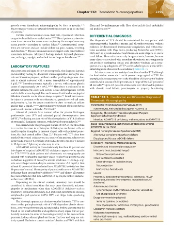
The symptoms and signs of TTP are nonspecific. The diagnosis depends ential diagnosis often must be considered (Table 132–1).
Schistocytes occur in a variety of conditions besides TTP, although
on laboratory testing to document microangiopathic hemolytic ane- the level seldom enters the 1 to 18 percent range typical of TTP. For
mia and thrombocytopenia, without another predisposing cause. Ane- example, schistocytes were seen in the blood film of 58 percent of healthy
mia is almost universal with a mean hemoglobin of approximately controls, with a mean of 0.05 percent and a range of 0 to 0.27 percent of
8 g/dL. 27,40 Thrombocytopenia typically is severe, with a mean platelet all red cells. Up to 0.6 percent schistocytes were observed in patients
42
count of approximately 20 × 10 /L. 26,27,40 Hemolysis is indicated by an with chronic renal failure, preeclampsia, or properly functioning
9
elevated reticulocyte count and serum lactate dehydrogenase (LDH),
undetectable serum haptoglobin, and increased total and unconjugated
bilirubin. Coombs test is almost always negative. Renal microvascu- TABLE 132–1. Classification and Differential Diagnosis of
7,8
lar injury is common with microhematuria, granular or red cell casts, Thrombotic Microangiopathy
and proteinuria, but the serum creatinine is often normal and seldom
greater than 2 mg/dL. 7,8,27,40 Approximately 50 percent of patients have a Thrombotic Thrombocytopenic Purpura (TTP)
positive anti-nuclear antibody (ANA) test. 40 Autoimmune, with antibodies against ADAMTS13
Almost all patients have normal values for plasma fibrinogen, Congenital Thrombotic Thrombocytopenic Purpura
prothrombin time (PT) and activated partial thromboplastin time (Upshaw-Schulman Syndrome)
(aPTT), reflecting a minor role of blood coagulation in TTP. Evidence Inherited ADAMTS13 deficiency, with mutations in ADAMTS13
7,8
of myocardial damage is common, with elevated troponin levels. 38,39
The characteristic morphological feature of TTP on the blood film Shiga Toxin-Producing Escherichia Coli Hemolytic Uremic
is a marked increase in schistocytes. Schistocytes are helmet cells, or Syndrome (STEC-HUS)
small irregular triangular or crescent shaped cells with pointed projec- Atypical Hemolytic Uremic Syndrome (aHUS)
tions, that lack central pallor (Chap. 2). Patients with TTP often have Alternative complement pathway defects
41
markedly increased schistocytes; in a study of six patients, schistocytes Diacylglycerol kinase ε (DGKE) defects
comprised a mean of 8.3 percent of all red cells with a range of 1 percent
to 18.4 percent. Spherocytes also may be seen. Secondary Thrombotic Microangiopathy
42
ADAMTS13 activity is characteristically less than 10 percent and Disseminated intravascular coagulation
this degree of acquired ADAMTS13 deficiency appears to be specific Infections (viral, bacterial, fungal)
for TTP. 13,14,43,44 If adult patients with thrombotic microangiopathy are Streptococcus pneumoniae
selected with no plausible secondary cause, no diarrheal prodrome, and Tissue transplant-associated
no features suggestive of hemolytic uremic syndrome (HUS) (e.g., olig-
uria, severe hypertension, dialysis, serum creatinine >3.5 mg/dL), then Chemotherapy or radiation injury
at least 80 percent of those selected have ADAMTS13 activity less than Tissue rejection
10 percent of normal. The majority of patients with severe ADAMTS13 Graft-versus-host disease
deficiency have autoantibody inhibitors 13,14,26,45 and almost all patients Cancer
have autoantibodies that bind ADAMTS13 by enzyme-linked immuno- Pregnancy associated (preeclampsia, eclampsia, HELLP
sorbent assay (ELISA). [hemolysis, elevated liver enzymes, low platelet count]
Depending on the clinical context, laboratory tests should be syndrome)
considered to detect conditions that may cause thrombotic microan- Autoimmune disorders
giopathy by mechanisms other than ADAMTS13 deficiency such as Systemic lupus erythematosus and other vasculitides
pregnancy, cobalamin deficiency, SLE and other autoimmune diseases,
antiphospholipid syndrome (APS), HIV, and Shiga toxin–producing Antiphospholipid syndrome
organisms. Drugs (commonly implicated)
The histologic appearance of microvascular lesions in TTP is con- Immune (quinine, ticlopidine)
sistent with a pathophysiologic role of VWF-dependent platelet throm- Toxic (cyclosporine, tacrolimus, mitomycin C, gemcitabine)
bosis. Amorphous thrombi and subendothelial hyaline deposits may be Cobalamin metabolic defects
found in the small arterioles and capillaries of any organ, but are par-
ticularly common (in order of decreasing severity) in the myocardium, Malignant hypertension
pancreas, kidney, adrenal gland and brain. The liver and lung are rela- Mechanical hemolysis (e.g., malfunctioning aortic or mitral
tively spared. The lesions consist mainly of platelets and VWF, with little valve prosthesis)
Kaushansky_chapter 132_p2253-2266.indd 2255 17/09/15 3:47 pm