
Page 2293 - Williams Hematology ( PDFDrive )
P. 2293
2268 Part XII: Hemostasis and Thrombosis Chapter 133: Venous Thrombosis 2269
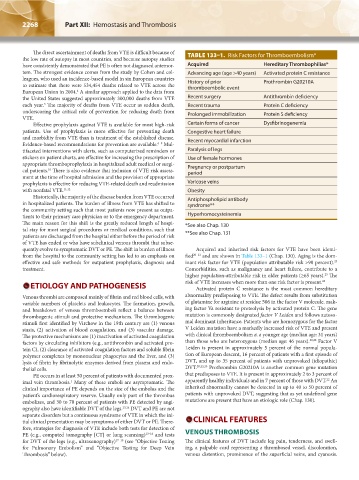
The direct ascertainment of deaths from VTE is difficult because of TABLE 133–1. Risk Factors for Thromboembolism*
the low rate of autopsy in most countries, and because autopsy studies
have consistently demonstrated that PE is often not diagnosed antemor- Acquired Hereditary Thrombophilias*
tem. The strongest evidence comes from the study by Cohen and col- Advancing age (age >40 years) Activated protein C resistance
leagues, who used an incidence-based model in six European countries History of prior Prothrombin G20210A
to estimate that there were 534,454 deaths related to VTE across the thromboembolic event
4
European Union in 2004. A similar approach applied to the data from
the United States suggested approximately 300,000 deaths from VTE Recent surgery Antithrombin deficiency
5
each year. The majority of deaths from VTE occur as sudden death, Recent trauma Protein C deficiency
underscoring the critical role of prevention for reducing death from Prolonged immobilization Protein S deficiency
VTE.
Effective prophylaxis against VTE is available for most high-risk Certain forms of cancer Dysfibrinogenemia
patients. Use of prophylaxis is more effective for preventing death Congestive heart failure
and morbidity from VTE than is treatment of the established disease. Recent myocardial infarction
6–9
Evidence-based recommendations for prevention are available. Mul-
tifaceted interventions with alerts, such as computerized reminders or Paralysis of legs
stickers on patient charts, are effective for increasing the prescription of Use of female hormones
appropriate thromboprophylaxis in hospitalized adult medical or surgi- Pregnancy or postpartum
cal patients. There is also evidence that inclusion of VTE risk assess- period
10
ment at the time of hospital admission and the provision of appropriate
prophylaxis is effective for reducing VTE-related death and readmission Varicose veins
with nonfatal VTE. 11,12 Obesity
Historically, the majority of the disease burden from VTE occurred Antiphospholipid antibody
in hospitalized patients. The burden of illness from VTE has shifted to syndrome**
the community setting such that most patients now present as outpa-
tients to their primary care physician or to the emergency department. Hyperhomocysteinemia
The main reason for this shift is the greatly reduced length of hospi- *See also Chap. 130
tal stay for most surgical procedures or medical conditions, such that
patients are discharged from the hospital either before the period of risk **See also Chap. 131
of VTE has ended or who have subclinical venous thrombi that subse-
quently evolve to symptomatic DVT or PE. The shift in burden of illness Acquired and inherited risk factors for VTE have been identi-
from the hospital to the community setting has led to an emphasis on fied 20–23 and are shown in Table 133–1 (Chap. 130). Aging is the dom-
effective and safe methods for outpatient prophylaxis, diagnosis and inant risk factor for VTE (population attributable risk >90 percent).
23
treatment. Comorbidities, such as malignancy and heart failure, contribute to a
23
higher population-attributable risk in older patients (≥65 years). The
ETIOLOGY AND PATHOGENESIS risk of VTE increases when more than one risk factor is present. 24
Activated protein C resistance is the most common hereditary
Venous thrombi are composed mainly of fibrin and red blood cells, with abnormality predisposing to VTE. The defect results from substitution
variable numbers of platelets and leukocytes. The formation, growth, of glutamine for arginine at residue 506 in the factor V molecule, mak-
and breakdown of venous thromboemboli reflect a balance between ing factor Va resistant to proteolysis by activated protein C. The gene
thrombogenic stimuli and protective mechanisms. The thrombogenic mutation is commonly designated factor V Leiden and follows autoso-
stimuli first identified by Virchow in the 19th century are (1) venous mal dominant inheritance. Patients who are homozygous for the factor
stasis, (2) activation of blood coagulation, and (3) vascular damage. V Leiden mutation have a markedly increased risk of VTE and present
The protective mechanisms are (1) inactivation of activated coagulation with clinical thromboembolism at a younger age (median age: 31 years)
factors by circulating inhibitors (e.g., antithrombin and activated pro- than those who are heterozygous (median age: 46 years). 20,22 Factor V
tein C), (2) clearance of activated coagulation factors and soluble fibrin Leiden is present in approximately 5 percent of the normal popula-
polymer complexes by mononuclear phagocytes and the liver, and (3) tion of European descent, 16 percent of patients with a first episode of
lysis of fibrin by fibrinolytic enzymes derived from plasma and endo- DVT, and up to 35 percent of patients with unprovoked (idiopathic)
thelial cells. DVT. 20,22,25 Prothrombin G20210A is another common gene mutation
PE occurs in at least 50 percent of patients with documented prox- that predisposes to VTE. It is present in approximately 2 to 3 percent of
22
1
imal vein thrombosis. Many of these emboli are asymptomatic. The apparently healthy individuals and in 7 percent of those with DVT. An
clinical importance of PE depends on the size of the embolus and the inherited abnormality cannot be detected in up to 40 to 50 percent of
patient’s cardiorespiratory reserve. Usually only part of the thrombus patients with unprovoked DVT, suggesting that as yet undefined gene
embolizes, and 30 to 70 percent of patients with PE detected by angi- mutations are present that have an etiologic role (Chap. 130).
ography also have identifiable DVT of the legs. 13,14 DVT and PE are not
separate disorders but a continuous syndrome of VTE in which the ini-
tial clinical presentation may be symptoms of either DVT or PE. There- CLINICAL FEATURES
fore, strategies for diagnosis of VTE include both tests for detection of
PE (e.g., computed tomography [CT] or lung scanning) 13–16 and tests VENOUS THROMBOSIS
for DVT of the legs (e.g., ultrasonography) 17–19 (see “Objective Testing The clinical features of DVT include leg pain, tenderness, and swell-
for Pulmonary Embolism” and “Objective Testing for Deep Vein ing, a palpable cord representing a thrombosed vessel, discoloration,
Thrombosis” below). venous distention, prominence of the superficial veins, and cyanosis.
Kaushansky_chapter 133_p2267-2280.indd 2268 9/18/15 10:52 AM