
Page 2297 - Williams Hematology ( PDFDrive )
P. 2297
2272 Part XII: Hemostasis and Thrombosis Chapter 133: Venous Thrombosis 2273
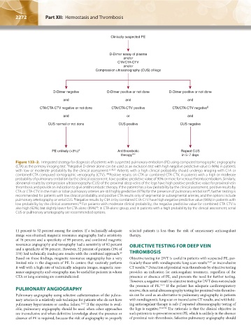
Clinically suspected PE
D-Dimer assay of plasma
and/or
CTA/CTA-CTV
and/or
Compression ultrasonography (CUS) of legs
D-Dimer negative D-Dimer positive or not done D-Dimer positive or not done
and and and
CTA/CTA-CTV negative or not done CTA/CTA-CTV positive CTA/CTA-CTV negative ‡
and or and
CUS normal or not done CUS positive CUS negative
PE unlikely (<3%) ∗ Antithrombotic Repeat CUS
therapy ∗∗ in 5–7 days
Figure 133–2. Integrated strategy for diagnosis of patients with suspected pulmonary embolism (PE) using computed tomographic angiography
(CTA) as the primary imaging test. *Negative D-dimer alone can be used as an exclusion test with high negative predictive value (>96%) in patients
with low or moderate probability by the clinical assessment. 27,30,31 Patients with a high clinical probability should undergo imaging with CTA or
combined CTA-computed tomographic venography (CTV). **Positive results on CTA or combined CTA-CTV, in patients with a high or moderate
probability of pulmonary embolism by the clinical assessment, have positive predictive value of 90% or more for venous thromboembolism. Similarly,
abnormal results by compression ultrasonography (CUS) of the proximal deep veins of the legs have high positive predictive value for proximal vein
thrombosis and provide an indication to give antithrombotic therapy. If the patient has a low probability by the clinical assessment, positive results by
43
CTA or CTA-CTV in the main or lobar pulmonary arteries are still highly predictive (97%) for the presence of pulmonary embolism ; further testing is
recommended for patients with low clinical probability and positive CTA results only of segmental or subsegmental arteries, and the options include
‡
pulmonary arteriography or serial CUS. Negative results by CTA or by combined CTA-CTV have high negative predictive value (96%) in patients with
43
low probability by the clinical assessment. For patients with moderate clinical probability, the negative predictive value for combined CTA-CTV is
43
also high (92%), but slightly lower for CTA alone (89%) ; in CTA-alone group, and in patients with a high probability by the clinical assessment, serial
CUS or pulmonary arteriography are recommended options.
11 percent to 52 percent among the centers. If a technically adequate selected patients is less than the risk of unnecessary anticoagulant
image was obtained, magnetic resonance angiography had a sensitivity therapy.
of 78 percent and a specificity of 99 percent, and combined magnetic
resonance angiography and venography had a sensitivity of 92 percent OBJECTIVE TESTING FOR DEEP VEIN
and a specificity of 96 percent. However, 52 percent of patients (194 of
370) had technically inadequate results with the combined approach. THROMBOSIS
49
Based on these findings, magnetic resonance angiography has a very Objective testing for DVT is useful in patients with suspected PE, par-
limited role in the diagnosis of PE. In centers that routinely perform ticularly those with nondiagnostic lung scan results 33,47 or inconclusive
it well with a high rate of technically adequate images, magnetic reso- CT results. Detection of proximal vein thrombosis by objective testing
50
nance angiography and venography may be useful for patients in whom provides an indication for anticoagulant treatment, regardless of the
CTA or lung scanning are contraindicated. presence or absence of PE, and prevents the need for further testing.
However, a negative result by objective testing for DVT does not exclude
the presence of PE. 13,14 If the patient has adequate cardiorespiratory
PULMONARY ANGIOGRAPHY reserve, then serial ultrasonography testing for proximal vein thrombo-
Pulmonary angiography using selective catheterization of the pulmo- sis can be used as an alternative to pulmonary angiography in patients
nary arteries is a relatively safe technique for patients who do not have with nondiagnostic lung scan or inconclusive CT results, and withhold-
pulmonary hypertension or cardiac failure. 13,15 If the expertise is avail- ing anticoagulant therapy is safe if repeated ultrasonography testing of
able, pulmonary angiography should be used when other approaches the legs is negative. 33,47,50 The rationale is that the clinical objective in
are inconclusive and when definitive knowledge about the presence or such patients is to prevent recurrent PE, which is unlikely in the absence
absence of PE is required, because the risk of angiography in properly of proximal vein thrombosis. Selective pulmonary angiography should
Kaushansky_chapter 133_p2267-2280.indd 2272 9/18/15 10:52 AM