
Page 2307 - Williams Hematology ( PDFDrive )
P. 2307
2282 Part XII: Hemostasis and Thrombosis Chapter 134: Atherothrombosis: Disease Initiation, Progression, and Treatment 2283
TABLE 134–1. Cardiovascular Risk Factors That Cause dysfunction are intimately connected and seminal to the initiation and
progression of atherosclerosis. Endothelial dysfunction occurs early in
Impaired Endothelium-Dependent Vasodilation
the development of plaque and is systemic in nature, afflicting vessels
Smoking throughout the arterial circulation without gross evidence of atheroscle-
Dyslipidemia rotic plaque formation. Emerging data indicate that proatherosclerotic
Hypertension genes are upregulated and antiatherosclerotic genes are downregulated
21
Diabetes mellitus in areas of turbulent blood flow, as seen at branch points of arteries,
resulting in vascular adhesion molecule expression and recruitment of
Hyperhomocysteinemia
monocytes. The atherosclerotic plaque initially may expand outward
22
rather than inward into the vessel wall, making some significant lesions
difficult to visualize by angiography. The components of the mature ath-
Cardiovascular morbidity and mortality is also recognized to be erosclerotic lesion include smooth muscle cells, macrophages, T lym-
23
exceedingly high in patients with chronic renal failure. 15,16 Increased phocytes, and calcification, in addition to accumulation of lipoproteins.
risk of premature atherosclerotic cardiovascular disease in patients Neutrophils and mast cells also are implicated in the atherosclerotic
22
on chronic hemodialysis has been known for many years, but recent process. Later in the process, increased activity of matrix metallo-
studies point to an increased risk even at early stages of chronic kidney proteinases in the atherosclerotic cap predisposes to plaque rupture or
diseases. Low glomerular filtration rates and/or proteinuria are inde- ulceration, resulting in tissue factor (TF) exposure and platelet adhe-
24
pendently associated with increased rates of cardiovascular disease. sion, culminating in thrombus formation. The thrombus may undergo
17
Other factors, such as sympathetic overactivity, are likely to contribute endogenous fibrinolysis with plaque healing or become occlusive and
18
to the pathophysiology of cardiac risk in these patients. Among other produce organ damage (e.g., myocardial infarction [MI]). In severe
emerging risk factors is obstructive sleep apnea, in which treatment may lesions, lamellar bone, presumably from endochondral calcification, may
25
improve cardiovascular outcomes. 19 appear. There is evolving evidence that extracellular vesicles (EVs), also
26
known as microparticles, are involved in the atherosclerotic process.
The following sections describe in detail the major manifestations of
ENDOTHELIAL DYSFUNCTION endothelial dysfunction that occur early in the atherosclerotic process.
Cardiovascular risk factors and abnormal blood rheology are thought
to result in endothelial dysfunction that predisposes the aorta and arter- Abnormal Vascular Tone
ies to atherosclerotic plaque development, sparing the arterioles and The importance of the endothelium in maintaining vascular tone
capillaries (Fig. 134–1). Endothelial dysfunction is a term that encom- was first recognized when endothelial cells of rabbit aorta were inad-
passes perturbations in the diverse physiologic functions of normal vertently removed and resulted in paradoxical vasoconstriction after
arteries, including regulation of vascular tone, inflammation, growth, administration of acetylcholine. The major endothelium-dependent
27
and preservation of blood fluidity. Lipid accumulation and endothelial vasodilator normally produced was found to be nitric oxide (NO), a free
20
Atherothrombosis: A generalized
and progressive process
Fatty Intermediate Fibrous Complicated Unstable
Foam cells streak lesion Atheroma plaque lesion/rupture angina
ACS
MI
Ischemic
stroke/TIA
Acute limb
ischemia
Clinically silent
Stable angina Cardiovascular
Increasing age intermittent claudication death
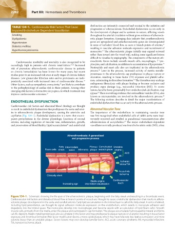
Figure 134–1. Schematic showing the life span of the atherosclerotic plaque, beginning with the fatty streak and resulting in a thrombotic event.
Cardiovascular risk factors and disturbed blood flow at branch points of vessels are thought to cause endothelial dysfunction that results in athero-
sclerotic plaque development in the aorta and conduit arteries. Early lipid accumulation in the intimal layer is called the fatty streak. A series of stimuli,
including lipid peroxidation, are thought to signal adhesion molecule expression on the endothelium, which results in monocyte adhesion and
diapedesis into the intimal space. The monocytes develop into macrophages and become sessile with accumulation of lipid (foam cells). Smooth
muscle cells, primarily from the media, enter the plaque and participate in cap formation. The plaque accumulates hydroxyapatite mineral and forms
calcific deposits. Matrix metalloproteinases also accumulate in the lesion and may predispose to plaque rupture or ulceration resulting in tissue factor
exposure and thrombus formation. Risk factor modification favors a more stable plaque, which may have relatively less lipid accumulation and more
sclerotic tissue than an unstable plaque. Severe lesions may even develop lamellar bone. ACS, acute coronary syndrome; MI, myocardial infarction;
TIA, transient ischemic attack.
Kaushansky_chapter 134_p2281-2302.indd 2282 17/09/15 3:49 pm