
Page 2383 - Williams Hematology ( PDFDrive )
P. 2383
2354 Part XIII: Transfusion Medicine Chapter 137: Human Leukocyte and Platelet Antigens 2355
TABLE 137–1. Number of Known Alleles for Each Human acids) are accommodated. 13,14 Class II antigens have a more restricted
tissue distribution, being found primarily on B lymphocytes and other
Leukocyte Antigen Locus as of July 2014
antigen-presenting cells such as dendritic cells, monocytes, and macro-
HLA CLASS I phages. They may also be expressed on activated endothelial cells and
Gene A B C E F G T lymphocytes. 12
The extraordinarily polymorphic nature of HLA has probably
Alleles 2884 3590 2375 15 22 50
evolved because of the need to present a very large array of different
HLA CLASS II antigenic peptides in host defense. Antigen processing and presenta-
Gene DRA DRB DQA DQB DPA DPB tion is a tightly regulated process, especially among the professional
antigen-presenting cells such as dendritic cells. A number of alternative
Alleles 7 1642 52 664 38 422
mechanisms have been demonstrated in vitro, such as cross-presentation,
NON-HLA whereby dendritic cells transfer antigen derived from endocytic sources
15
Gene MICA MICB TAP1 TAP2 to the class I pathway, but are poorly understood. One promising area
of research is the ability of HLA molecules to present antigenic peptides
Alleles 100 40 12 12
derived from tumors. Such peptides could arise via point mutation,
HLA, human leukocyte antigen (HLA). or reactivation of a normally silent gene that produces a peptide that
Data from Robinson J, Waller MJ, Parham P, et al: IMGT/HLA and IMGT/ can bind to HLA and induce a T-cell immune response. Several such
MHC: Sequence databases for the study of the major histocompati- peptides (melanoma-associated gene [MAGE] antigens) have been
bility complex. Nucleic Acids Res 31(1):311–314, 2003. identified for melanoma. 16
and the light β chain (Mr 29,000) are encoded in the MHC region. Class II NOMENCLATURE
molecules, like class I, consist of an extracellular hydrophilic NH – Distinguishing polymorphic variations among HLA antigens is clin-
2
terminal region, a hydrophobic transmembrane region, and an intracel- ically important in stem cell transplantation. Terminology used to
lular COOH– terminus region. Unlike class I antigens, the extracellular describe accepted HLA alleles or antigens is standardized by the
regions of each chain contain only two domains. The two domains of World Health Organization (WHO), Nomenclature Committee for
the α chain are designated α and α , and the two domains of the β-chain Factors of the HLA System, which issues biannual reports and monthly
2
1
are called β and β . The α chain of HLA-DR is constant for all HLA-DR updates. In addition, an HLA dictionary defining HLA antigens, their
4
2
1
molecules, whereas the β chain is polymorphic and determines spec- assigned nomenclature, and serologic equivalents is published period-
ificity of the molecule. Both α and β chains of HLA-DQ and -DP are ically. The nomenclature committee approved major changes to the
5
polymorphic, although the β chain is more so than the α chain. In all system that were implemented in 2010. The revisions were designed to
17
class II antigens the β domain represents the most polymorphic region. accommodate the unexpected number of new sequenced alleles. Under
1
The structure of HLA-DR is essentially similar to the structure of class I this system, colons are used as delimiters to separate fields. The first field
molecules. Class II antigens present peptides from exogenous sources, signifies the allele family that often corresponds to the serological antigen.
such as bacterial pathogens, to CD4+ cells. The binding groove is more The second field denotes the alleles, assigned in order of determination.
open than that of class I, and peptides of longer length (11 to 18 amino The third field is used for defining synonymous nucleotide substitutions.
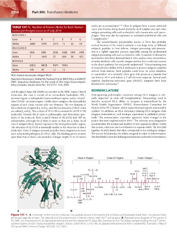
Figure 137–1. A. Schematic of the HLA-A2 molecule. The peptide groove is formed by the α helices and β-pleated sheet floor. The groove holds
processed peptide antigen. The peptide and the polymorphic α helices interact with the T-cell receptor. B. Representative diagram of the genes of
the MHC on chromosome 6. (A, reproduced with permission from Bjorkman PJ, Saper MA, Samraoui B, et al: The foreign antigen binding site and T cell rec-
ognition regions of class I histocompatibility antigens. Nature 329(6139):512–518, 1987. B, adapted with permission from Campbell RD, Trowsdale J: Map of
the human MHC, Immunol Today 14(7):349–352, 1993.)
Kaushansky_chapter 137_p2353-2364.indd 2354 9/21/15 3:49 PM