
Page 333 - Williams Hematology ( PDFDrive )
P. 333
308 Part IV: Molecular and Cellular Hematology Chapter 21: Dendritic Cells and Adaptive Immunity 309
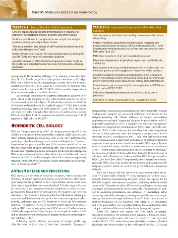
TABLE 21–1. Role of Dendritic Cell in Immunity TABLE 21–2. Important Components of Dendritic Cell
Sensors: rapid and appropriate differentiation in response to Function
pathogen-associated molecular patterns and other signals Cell processes or dendrites and motility: numerous and continu-
Sentinels: positioned in peripheral tissues to optimize antigen ally probing
capture and migrate to lymphoid tissues Antigen handling: specialized antigen uptake receptors and
Tolerance: deletion and anergy of self-reactive lymphocytes and processing pathways for classic (MHC) and nonclassic (CD1 and
induction of regulatory T cells others) presenting molecules, including cross-presentation onto
MHC class I and CD1
Innate resistance: activation of innate lymphocytes, including NK
and NKT cells, secretion of protective cytokines MHC class II products: high and regulated expression
Adaptive immunity: differentiation of quiescent, naïve T cells to Migration in lymphatics to lymphoid organs and localization to
form effectors, establishment of memory lymphocytes, antibody T-cell areas
responses Environmental sensing: multiple receptors for microbial and non-
microbial products and exaggerated responses to the products
appropriate to the invading pathogen. For example, under the influ- Cytokine receptors, including hematopoietins (flt3L and granu-
13
ence of DCs, T cells can preferentially produce interferon-γ (T-helper locyte-macrophage colony-stimulating factor, but not monocyte
colony-stimulating factor, granulocyte colony-stimulating factor)
[Th] type 1 cells) to activate macrophages to resist infection by intra-
cellular microbes; or IL-4, IL-5, and IL-13 (Th2 cells) to mobilize white Chemokine receptors, especially for homing to tissues (CCR6) and
cells to resist helminths; or IL-17 (Th17 cells) to mobilize phagocytes at lymph nodes (CCR7, CCR2)
body surfaces to resist extracellular bacteria. Induction of peripheral tolerance via intrinsic and extrinsic
An essential counterpart to adaptive immunity is adaptive toler- pathways
ance, which is the silencing of cells with receptors reactive to self or Activation of innate lymphocytes (e.g., natural killer cells)
harmless environmental antigens. T cells develop tolerance centrally in
the thymus and peripherally in lymphoid organs. 14,15 DCs play a role in
inducing tolerance, especially with regard to T cells. DCs can silence
self-reactive T cells, through either deletion or functional inactivation. antigens refer to molecules processed directly following uptake, whereas
DCs can also drive T cells to suppress immunity by expressing IL-10 (T “endogenous” antigens are processed following biosynthesis in the
regulatory [Tr]1 cells) or FOXP3. 16 antigen-presenting cell. Classic pathways of antigen presentation
emphasize processing of “exogenous” antigens for presentation on MHC
DENDRITIC CELL BIOLOGY II–peptide complexes to CD4+ T lymphocytes, whereas “endogenous”
antigens were targeted for presentation on MHC class I–peptide com-
DCs are “antigen-presenting cells.” An antigen-presenting cell is any plexes to CD8+ T cells. However, it is now clear that there is significant
cell that uses its major histocompatibility complex (MHC) products (or overlap in these pathways such that exogenous antigens may also be
other antigen-presenting molecules, such as the CD1 molecules that presented on MHC I products to CD8+ T cells. This pathway is termed
present glycolipids and lipoglycans) to bind and display (i.e., “present”) cross-presentation and is important for initiation of antitumor immune
fragments of antigen to lymphocytes. DCs are more specialized or pro- responses. Cross-presentation is well developed in DCs, especially those
fessional than other antigen-presenting cells. This is because DCs have found in lymphoid tissues, and leads to either tolerance or activation of
23
efficient and regulated pathways for antigen uptake and processing, and CD8+ T lymphocytes, depending upon the DC maturation stimulus.
DCs possess dozens of features that allow them to initiate and control For instance, products of dying cells, from transplants, tumors, foci of
immunity (Table 21–2). For example, when DCs mature in response to infection, and self-tissues are endocytosed by DCs then presented on
infection, hundreds, even thousands, of gene transcripts can be upregu- MHC Class I to CD8+ cells. 24,25 Importantly, cross-presentation of anti-
lated or downregulated. 17,18 gens onto MHC class I can involve the proteasome and transporters for
antigenic peptides, which are used in the presentation of endogenous
ANTIGEN UPTAKE AND PROCESSING antigens.
DCs are a major cell type involved in cross-presentation of pro-
DCs express a wide array of endocytic receptors, which enhance the teins, 26,27 and probably of lipids. 28,29 Cross-presentation has been docu-
efficiency of antigen capture, processing, and presentation. Many endo- mented involving nonreplicating microbes, dying cells, ligands for the
cytic receptors are predicted to be C-type lectins, and in some cases DEC205 receptor, and immune complexes, including antibody-coated
their natural ligands have not been identified. DCs also express Fcγ and tumor cells. This pathway allows DCs to induce tolerance or immunity
Fcε receptors, which recognize immune complexes, as well as scaven- to antigens not synthesized de novo in these cells. Fcγ receptors, in addi-
ger receptors. Recognition of pathogens by DC receptors can have two tion to mediating presentation, can influence DC maturation, either
outcomes. One outcome is immune activation followed by antigen pre- enhancing maturation through activating forms of the receptor or pre-
sentation and development of a productive immune responses. Alter- venting maturation through inhibitory forms. Such consequences of
30
natively, pathogens may use DC receptors to evade the host immune antibody binding to DC Fc receptors, with regard to DC maturation
response. For example, DC-SIGN (CD209) a lectin expressed on DCs, is and cross-presentation, are important features for consideration when
used by HIV-1 and cytomegalovirus (CMV) to reach T cells and endo- trying to understand the use of antibodies as therapeutic agents.
thelial cells, respectively 19,20 ; by Dengue virus to replicate within DCs ; Some DCs subsets also express the CD1 family of antigen-
21
and by Mycobacterium tuberculosis to trigger production of the suppres- presenting molecules. For example, CD1a typically is found on epider-
sive cytokine IL-10. 22 mal Langerhans cells in skin, whereas CD1b and CD1c are expressed
Following uptake, efficient processing of antigen yields pep- on dermal DCs. CD1 molecules present glycolipids, whereas microbial
tides that bind to MHC class II and class I products. “Exogenous” glycolipids are the best studied to date with regard to CD1a, CD1b, and
Kaushansky_chapter 21_p0307-0312.indd 308 9/17/15 5:52 PM