
Page 698 - Williams Hematology ( PDFDrive )
P. 698
672 Part VI: The Erythrocyte Chapter 46: Erythrocyte Membrane Disorders 673
+
influx activates the Na -K ATPase cation pump, which increases ATP and this is the primary cause of the chronic hemolysis experienced by
+
turnover and glycolysis. Spherocytes are dehydrated, especially cells HS patients (see Fig. 46–12). The reduced deformability of spherocytes
obtained from the splenic pulp, but the underlying mechanism has not impedes their passage through the interendothelial slits separating the
been clearly defined. The acidic environment of the spleen and oxida- splenic cords of the red pulp from the splenic sinuses. The decrease in
–
+
tive damage by splenic macrophages increase the activity of the K Cl red cell deformability is primarily related to a loss of surface area and, to
cotransporter, which may play a role in dehydration. The hyperactive a lesser extent, to an increase in internal viscosity as a result of mild cel-
+
+
Na -K ATPase pump may also contribute as three sodium ions are lular dehydration. Ex vivo experiments using perfused human spleens
extruded in exchange for two potassium ions, and this loss of monova- and red cells treated with lysophosphatidylcholine to induce spherocy-
lent cations is accompanied by the loss of water. Dehydration may also tosis revealed that the degree of splenic retention correlated with the
be related to loss of surface area. reduction in the surface-area-to-volume ratio. 99
The spleen is a metabolically hostile environment with a decreased
Molecular Determinants of Clinical Severity pH, low concentrations of glucose and ATP, and increased oxidants, all
Affected individuals of the same kindred typically experience similar of which are detrimental to the red cell. Spherocytes are “conditioned”
degrees of hemolysis. However, in some families the clinical expres- during erythrostasis in the spleen and become more osmotically fragile
sion is variable and this may be influenced by several factors. Low- and increasingly spherocytic. Exposure to macrophages in the spleen
100
expression alleles decrease transcription of the gene or influence the eventually leads to erythrophagocytosis and destruction.
expression or incorporation of the protein into the membrane, but there
is no phenotypic effect in the heterozygous state because the normal Inheritance
allele compensates for the deleterious effect. However, when inherited In approximately 75 percent of HS patients, inheritance is autosomal
with a mutant allele that causes HS, it exacerbates the clinical expres- dominant. In the remaining patients, the disorder may be autosomal
sion of the disease. Examples of low-expression alleles that influence recessive or result from de novo mutations, which is relatively com-
HS include band 3 Genas, band 3 Mondego, and two α-spectrin alleles, mon. 101,102 Mutations in α-spectrin or protein 4.2 are often associated
α LELY and α LEPRA 77,81,91–94 with recessive HS.
.
Variable penetrance of the defective gene, a de novo mutation or
a mild form of recessively inherited HS may also influence the clini- Clinical Features
cal severity. Double heterozygosity for two mild band 3 mutations can The clinical manifestations of HS vary widely. The typical clinical pic-
76
have an additive effect and rare cases caused by homozygous defects ture combines evidence of hemolysis (anemia, jaundice, reticulocytosis,
in band 3 result in severe transfusion-dependent hemolytic anemia gallstones, splenomegaly) with spherocytosis (spherocytes on the blood
or fetal death. 91,95,96 Coinheritance of other hematologic disorders or film and increased osmotic fragility) and a positive family history. Mild,
Gilbert syndrome, caused by homozygosity for a polymorphism in the moderate, and severe forms of HS have been defined according to dif-
promoter of the uridine diphosphate-glucuronosyltransferase (UGT1) ferences in hemoglobin, bilirubin, and reticulocyte counts (Table 46–3),
gene, can also alter the clinical symptoms. 63,97,98 which can be correlated with the degree of compensation for hemolysis.
Initial assessment of a patient with suspected HS should include a fam-
Role of the Spleen ily history and questions about history of anemia, jaundice, gallstones,
The spleen plays a secondary but important role in the pathophysiology and splenectomy. Physical examination should seek signs such as scleral
of HS. Spherocytes are retained and ultimately destroyed in the spleen icterus, jaundice, and splenomegaly.
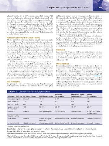
TABLE 46–3. Classification of Hereditary Spherocytosis
Moderate Moderately Severe Severe
Laboratory Findings HS Trait or Carrier Mild Spherocytosis Spherocytosis Spherocytosis* Spherocytosis †
Hemoglobin (g/dL) Normal 11–15 8–12 6–8 <6
Reticulocytes (%) 1–2 3–8 ± 8 ≥10 ≥10
Bilirubin (mg/dL) 0–1 1–2 ± 2 2–3 ≥3
Spectrin content 100 80–100 50–80 40–80 § 20–50
(% of normal) ‡
Blood film Normal Mild spherocytosis Spherocytosis Spherocytosis Spherocytosis and
poikilocytosis
Osmotic fragility
Fresh blood Normal Normal or slightly Distinctly increased Distinctly increased Distinctly increased
increased
Incubated blood Slightly increased Distinctly increased Distinctly increased Distinctly increased Markedly increased
*Values in untransfused patients.
† By definition, patients with severe spherocytosis are transfusion-dependent. Values were obtained immediately prior to transfusion.
‡ Normal: 245 ± 27 × 10 spectrin dimers per erythrocyte.
3
§ Spectrin content is variable in this group of patients, presumably reflecting heterogeneity of the underlying pathophysiology.
Adapted with permission from Eber SW, Armbrust R, Schröter W: Variable clinical severity of hereditary spherocytosis: Relation to erythrocytic
spectrin concentration, osmotic fragility, and autohemolysis. J Pediatr 1990 Sep;117(3):409–416.
Kaushansky_chapter 46_p0661-0688.indd 673 9/17/15 6:42 PM