
Page 731 - Williams Hematology ( PDFDrive )
P. 731
706 Part VI: The Erythrocyte Chapter 47: Erythrocyte Enzyme Disorders 707
reported missense mutations affect residues of the catalytic site, suggest-
ing that the reduced catalytic efficiency and/or instability result from
secondary effects related to conformational changes. 248
Acquired deficiency of P5′N1 may result from lead poisoning.
2+
Structural studies have shown that Pb specifically binds within the
2+
active site, in a different position than Mg but with much higher affin-
248
ity. Because simultaneous binding of Mg and Pb is not possible,
2+
2+
Pb outcompetes Mg , thereby preventing this essential cofactor from
2+
2+
binding, thus abolishing catalytic activity. P5′N1 activity is also inhib-
ited in β-thalassemia and related disorders that result in excess α-globin
chains, such as hemoglobin E, probably from oxidative damage induced
by excess α-globin chains. 436,437
MECHANISM OF HEMOLYSIS
G6PD Deficiency
The life span of G6PD-deficient red cells is shortened under many cir-
cumstances, particularly during drug administration and infection. The
exact reason for this is not known.
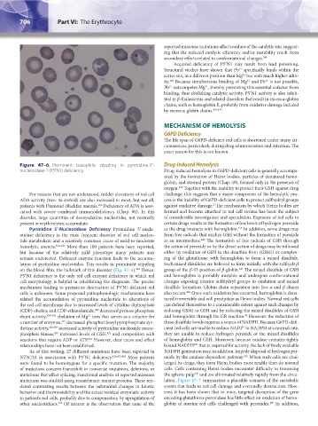
Figure 47–6. Prominent basophilic stippling in pyrimidine-5′- Drug-Induced Hemolysis
nucleotidase-1 (P5′N1) deficiency. Drug-induced hemolysis in G6PD-deficient cells is generally accompa-
nied by the formation of Heinz bodies, particles of denatured hemo-
globin, and stromal protein (Chap. 49), formed only in the presence of
oxygen. Together with the inability to protect their GSH against drug
438
For reasons that are not understood, milder elevations of red cell challenge, this suggests that a major component of the hemolytic pro-
ADA activity (two- to sixfold) are also increased in most, but not all, cess is the inability of G6PD-deficient cells to protect sulfhydryl groups
2
186
patients with Diamond-Blackfan anemia. Deficiency of ADA is asso- against oxidative damage. The mechanism by which Heinz bodies are
ciated with severe combined immunodeficiency (Chap. 80). In this formed and become attached to red cell stroma has been the subject
disorder, large quantities of deoxyadenine nucleotides, not normally of considerable investigation and speculation. Exposure of red cells to
present in erythrocytes, accumulate. certain drugs results in the formation of low levels of hydrogen peroxide
439
Pyrimidine 5′-Nucleotidase Deficiency Pyrimidine 5′-nucle- as the drug interacts with hemoglobin. In addition, some drugs may
otidase deficiency is the most frequent disorder of red cell nucleo- form free radicals that oxidize GSH without the formation of peroxide
440
tide metabolism and a relatively common cause of mild-to-moderate as an intermediate. The formation of free radicals of GSH through
hemolytic anemia. 420–422 More than 100 patients have been reported, the action of peroxide or by the direct action of drugs may be followed
but because of the relatively mild phenotype many patients may either by oxidation of GSH to the disulfide form (GSSG) or complex-
remain undetected. Deficient enzyme function leads to the accumu- ing of the glutathione with hemoglobin to form a mixed disulfide.
lation of pyrimidine nucleotides. This results in prominent stippling Such mixed disulfides are believed to form initially with the sulfhydryl
244
on the blood film, the hallmark of this disorder (Fig. 47–6). Hence, group of the β-93 position of β-globin. The mixed disulfide of GSH
441
P5′N1 deficiency is the only red cell enzyme deficiency in which red and hemoglobin is probably unstable and undergoes conformational
cell morphology is helpful in establishing the diagnosis. The precise changes exposing interior sulfhydryl groups to oxidation and mixed
mechanism leading to premature destruction of P5′N1-deficient red disulfide formation. Globin chain separation into free α and β chains
cells is unknown. Some proposed pathophysiologic mechanisms have also occurs. Once such oxidation has occurred, hemoglobin is dena-
442
related the accumulation of pyrimidine nucleotide to alterations of tured irreversibly and will precipitate as Heinz bodies. Normal red cells
the red cell membrane due to increased levels of cytidine diphosphate can defend themselves to a considerable extent against such changes by
423
(CDP)-choline and CDP-ethanolamide, decreased pentose phosphate reducing GSSG to GSH and by reducing the mixed disulfides of GSH
42
2+
shunt activity, 424–426 chelation of Mg ions that serves as a cofactor for and hemoglobin through the GR reaction. However, the reduction of
a number of enzymes, decreased phosphoribosyl pyrophosphate syn- these disulfide bonds requires a source of NADPH. Because G6PD-defi-
427
+
thetase activity, 428,429 increased activity of pyrimidine nucleoside mono- cient red cells are unable to reduce NADP to NADPH at a normal rate,
phosphate kinase, increased levels of GSH, and competition with they are unable to reduce hydrogen peroxide or the mixed disulfides
430
431
reactions that require ADP or ATP. However, clear cause-and-effect of hemoglobin and GSH. Moreover, because catalase contains tightly
432
443
relationships have not been established. bound NADPH that is required for activity, the lack of freely available
As of this writing, 27 different mutations have been reported in NADPH generation may, in addition, impede disposal of hydrogen per-
NT5C3A in association with P5′N1 deficiency. 420,433,434 Most patients oxide by the catalase-dependent pathway. When such cells are chal-
444
were found to be homozygous for a specific mutation. The majority lenged by drugs, they form Heinz bodies more readily than do normal
of mutations concern frameshift or nonsense mutations, deletions, or cells. Cells containing Heinz bodies encounter difficulty in traversing
445
mutations that affect splicing. Functional analysis of reported missense the splenic pulp and are eliminated relatively rapidly from the circu-
mutations was studied using recombinant mutant proteins. These ren- lation. Figure 47–7 summarizes a plausible scenario of the metabolic
dered contrasting results between the substantial changes in kinetic events that leads to red cell damage and eventually destruction. How-
behavior and thermostability and the actual residual enzymatic activity ever, it has been shown that in mice, targeted disruption of the gene
in patient’s red cells, probably due to compensation by upregulation of encoding glutathione peroxidase has little effect on oxidation of hemo-
199
435
other nucleotidases. Of interest is the observation that none of the globin of murine red cells challenged with peroxides. In addition,
Kaushansky_chapter 47_p0689-0724.indd 706 9/17/15 6:44 PM