
Page 732 - Williams Hematology ( PDFDrive )
P. 732
706 Part VI: The Erythrocyte Chapter 47: Erythrocyte Enzyme Disorders 707
Sup Dismut of the acid phosphatase gene have been attributed to a decrease in the
–
O 2 f isoforms of this tyrosine phosphatase and, consequently, low GSH
levels. Immunologic factors do not seem to play a role in favism.
455
456
Increased levels of red cell calcium 457,458 and consequent “cross-
DRUG + HbO 2 H O 2 bonding” of membranes may occur. Other membrane alterations that
2
have been described are oxidation and clustering of membrane proteins,
hemichrome binding to the internal face of the membrane, destabiliza-
tion of the membrane, and the release of microvesicles. 459–462
GSH-Px
Icterus Neonatorum
G6PD-deficient neonates are at increased risk of developing severe ict-
GSSG GSH
erus neonatorum. The icterus is frequently unaccompanied by changes
in hematologic indices reflective of a hemolytic process. 463–465 The rea-
+ +
son for this discrepancy is unclear. Icterus probably results principally
NADPH GR NADP from inadequate processing of bilirubin by the immature liver of G6PD-
deficient infants. The demonstrated increase in carboxyhemoglobin
G-6-PD
levels, indicative of increased heme catabolism, suggests, however, that
shortening of red cell life span also plays a role. A predisposing factor
466
for severe jaundice in G6PD deficiency is mutation of the uridine diphos-
6–Phosphogluconate Glucose-6-P phoglucuronate glucuronosyltransferase-1 gene (UGT1A1) promoter,
467
468
or, in Asia, the c.211G>A coding mutation. In adults, these mutations
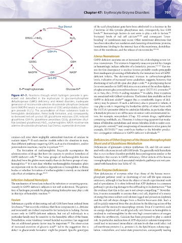
Figure 47–7. Reactions through which hydrogen peroxide is gen- are associated with Gilbert syndrome. The limited data available on liver
erated and detoxified in the erythrocyte. In glucose-6-phosphate G6PD in deficient adults suggest that a considerable degree of defi-
469
dehydrogenase (G6PD) deficiency and related disorders, inadequate ciency may be present. If such a deficiency also is present in infants, it
generation of nicotinamide adenine dinucleotide phosphate (reduced
form) (NADPH) results in accumulation of GSSG and probably of hydro- may play a role in impairing the borderline ability of infant livers with
gen peroxide (H O ). The accumulation of these substances leads to the UGT1A1 promoter defect to catabolize bilirubin, in particular when
2
2
hemoglobin denaturation, Heinz body formation, and, consequently, a hemolytic process is set off because of contact with environmental fac-
to decreased red cell survival. GR, glutathione reductase; GSH, reduced tors, for example, neocytolysis (Chap. 33) certain drugs, naphthalene
glutathione; GSH-Px, glutathione peroxidase; GSSG, glutathione disul- containing mothballs, etc. However, it is becoming apparent that modu-
fide (oxidized glutathione); HbO , oxyhemoglobin; NADP, nicotinamide lation of bilirubin metabolism and serum bilirubin levels is under com-
2
adenine dinucleotide phosphate; Sup Dismut, superoxide dismutase. plex genetic control, and coexpressing of mutations in other genes, for
470
471
example, SLCO1B3, may contribute further to the bilirubin produc-
tion-conjugation imbalance in G6PD-deficient individuals. 472
catalase-null mice show negligible antioxidant function of catalase in
oxidant injury. If such murine models reflect the situation in man, Deficiencies of Other Enzymes of the Hexose Monophosphate
446
then different pathways requiring GSH, such as the thioredoxin, and/or Shunt and of Glutathione Metabolism
peroxiredoxin reactions, may be important. 446,447 Deficiencies of glutamate cysteine synthetase, GS, and GR are associ-
The formation of methemoglobin frequently accompanies the ated with a decrease in red cell GSH levels. The generally mild hemolysis
administration of drugs that have the capacity to produce hemolysis of that occurs in these disorders probably has a pathogenesis similar to the
G6PD-deficient cells. The heme groups of methemoglobin become hemolysis that occurs in G6PD deficiency. Other defects of the hexose
448
detached from the globin more readily than do the heme groups of oxy- monophosphate shunt and associated metabolic pathways are not asso-
hemoglobin. It is not clear whether methemoglobin formation plays ciated with hemolysis (see Table 47–3).
449
an important role in the oxidative degradation of hemoglobin to Heinz
bodies or whether formation of methemoglobin is merely an incidental Other Enzyme Deficiencies
side effect of oxidative drugs. 450,451 How deficiencies of enzymes other than those of the hexose mono-
phosphate pathway result in shortening of red cell life-span remains
Infection-Induced Hemolysis unknown, although it has been the object of much experimental work
The mechanism of hemolysis induced by infection or occurring sponta- and of speculation. It is often believed that ATP depletion is a common
neously in G6PD-deficient subjects is not well understood. The genera- pathway in producing damage to the cell leading to its destruction, but
473
474
tion of hydrogen peroxide by phagocytizing leukocytes may play a role the evidence that this is the case is not always compelling. Neverthe-
in this type of hemolytic reaction. 451 less, it seems reasonable to assume that a red cell, deprived of a source
of energy becomes sodium and calcium logged and potassium depleted,
Favism and the red cell shape changes from a flexible biconcave disk. Such a
Substances capable of destroying red cell GSH have been isolated from cell is quickly removed from the circulation by the filtering action of the
452
fava beans, but scientific evidence that these components (i.e., divicine spleen and the monocyte-macrophage system. Even if it survived, such
and isouramil) are indeed responsible for hemolysis is lacking. Favism an energy-deprived cell would gradually turn brown as hemoglobin is
occurs only in G6PD-deficient subjects, but not all individuals in a oxidized to methemoglobin by the very high concentrations of oxygen
particular family may be sensitive to the hemolytic effect of the beans. within the erythrocyte. Calcium has been proposed to play a central
Nonetheless, some tendency toward familial occurrence has suggested role. In particular, malfunction of ATP-dependent calcium transporters
453
that an additional genetic factor may be important. The observation could lead to increased intracellular calcium levels that could affect red
454
of increased excretion of glucaric acid led to the suggestion that a cell membrane proteins (i.e., protein 4.1), the lipid bilayer, volume regu-
defect in glucuronide formation might be present. Specific genotypes lation, metabolism, and redox state preservation, consequently leading
Kaushansky_chapter 47_p0689-0724.indd 707 9/17/15 6:44 PM