
Page 830 - Williams Hematology ( PDFDrive )
P. 830
804 Part VI: The Erythrocyte Chapter 51: Fragmentation Hemolytic Anemia 805
A B
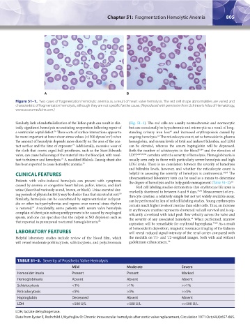
Figure 51–1. Two cases of fragmentation hemolytic anemia as a result of heart valve hemolysis. The red cell shape abnormalities are varied and
characteristic of fragmentation hemolysis, although they are not specific for the cause. (Reproduced with permission from Lichtman’s Atlas of Hematology,
www.accessmedicine.com.)
Similarly, lack of endothelialization of the Teflon patch can result in clin- (Fig. 51–1). The red cells are usually normochromic and normocytic
ically significant hemolysis necessitating reoperation following repair of but can occasionally be hypochromic and microcytic as a result of long-
a ventricular septal defect. These sorts of surface interactions appear to standing urinary iron loss and increased erythropoiesis caused by
61
76
be more important at lower shear-stress values (<1500 dynes/cm ) when ongoing hemolysis. The reticulocyte count, urine hemosiderin, plasma
2
62
the amount of hemolysis depends more directly on the area of the con- hemoglobin, and serum levels of total and indirect bilirubin, and LDH
tact surface and the time of exposure. Additionally, excessive wear of can be elevated, whereas the serum haptoglobin will be depressed.
77
the cloth that covers caged-ball prostheses, such as the Starr-Edwards Both the number of schistocytes in the blood 61,64 and the elevation of
valve, can cause ballooning of the material into the blood jet, with resul- LDH 64,65,83,84 correlate with the severity of hemolysis. Hemoglobinuria is
tant turbulence and hemolysis. A modified Blalock-Taussig shunt also usually seen only in those with particularly severe hemolysis and high
78
has been reported to cause hemolytic anemia. 79 LDH levels. There is no correlation between the severity of hemolysis
and bilirubin levels, however, and whether the reticulocyte count is
CLINICAL FEATURES helpful in assessing the severity of hemolysis is controversial. 64,65 The
Patients with valve-induced hemolysis can present with symptoms aforementioned laboratory tests can be used as a means to determine
the degree of hemolysis and to help guide management (Table 51–2).
64
caused by anemia or congestive heart failure, pallor, icterus, and dark Red cell labeling studies demonstrate that erythrocyte life span is
urine (described variously as red, brown, or black). Urine excreted dur- markedly shortened to between 6 and 9 days. 76,80 Measurement of ery-
80
ing periods of physical activity may be darker than that excreted at rest. throcyte creatine, a relatively simple but not yet widely available assay,
Similarly, hemolysis can be exacerbated by supraventricular tachycar- can be performed in lieu of red cell labeling studies. Young erythrocytes
dia or other tachyarrhythmias and regress once normal sinus rhythm contain much higher levels of creatine than older cells. Thus, an increase
is restored. Anecdotally, some patients with severe valve hemolysis in erythrocyte creatine represents shortened red cell survival and is sig-
81
complain of chest pain subsequently proven to be caused by esophageal nificantly correlated with total peak flow velocity across the valve and
spasm, and one can speculate that the culprit is NO depletion such as the severity of any associated hemolysis. When performed, marrow
85
that reported in paroxysmal nocturnal hemoglobinuria. 82 aspiration will be remarkable for erythroid hyperplasia. 75,80 As a result
of hemosiderin deposition, magnetic resonance imaging of the kidneys
LABORATORY FEATURES will reveal reduced signal intensity of the renal cortex compared with
Helpful laboratory studies include review of the blood film, which the medulla on T1- and T2-weighted images, both with and without
will reveal moderate poikilocytosis, schistocytosis, and polychromasia gadolinium enhancement. 86
TABLE 51–2. Severity of Prosthetic Valve Hemolysis
Mild Moderate Severe
Hemosider inuria Present Present Marked
Hemoglobinuria Absent Absent Absent
Schistocytosis <1% >1% >>1%
Reticulocytosis <5% >5% >>5%
Haptoglobin Decreased Absent Absent
LDH <500 U/L >500 U/L >>500 U/L
LDH, lactate dehydrogenase.
Data from Eyster E, Rothchild J, Mychajliw O: Chronic intravascular hemolysis after aortic valve replacement, Circulation 1971 Oct;44(4):657-665.
Kaushansky_chapter 51_p0801-0008.indd 805 9/17/15 2:42 PM