
Page 841 - Williams Hematology ( PDFDrive )
P. 841
816 Part VI: The Erythrocyte Chapter 53: Hemolytic Anemia Resulting from Infections with Microorganisms 817
64
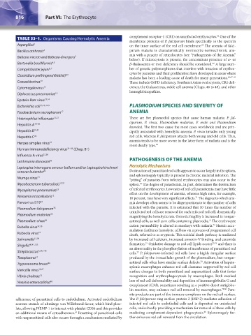
TABLE 53–1. Organisms Causing Hemolytic Anemia complement receptor-1 (CR1) on uninfected erythrocytes. One of the
membrane proteins of P. falciparum binds specifically to the spectrin
Aspergillus 1 on the inner surface of the red cell membrane. The anemia of falci-
65
Bacillus anthracis 2 parum malaria is characteristically normocytic-normochromic ane-
mia with a paucity of reticulocytes (see “Pathogenesis of the Anemia”
Babesia microti and Babesia divergens 3
below). If microcytosis is present, the concomitant presence of α- or
Bartonella bacilliformis 4,5 β-thalassemia or iron deficiency should be considered. A large num-
66
Campylobacter jejuni 6,7 ber of genetic polymorphisms that interfere with invasion of erythro-
cytes by parasites and their proliferation have developed in areas where
Clostridium perfringens(Welchii) 8,9
malaria has been a leading cause of death for many generations. 64,67–69
Coxsackievirus 10 These include G6PD deficiency, Southeast Asian ovalocytosis, CR1 defi-
Cytomegalovirus 11 ciency, the thalassemias, sickle cell anemia (Chaps. 46 to 48), and other
hemoglobinopathies.
Diplococcus pneumoniae 12
Epstein-Barr virus 13,14
Escherichia coli 15,16, 123 PLASMODIUM SPECIES AND SEVERITY OF
Fusobacterium necrophorum 17 ANEMIA
Haemophilus influenzae 12,23 There are five plasmodial species that cause human malaria: P. fal-
ciparum, P. vivax, Plasmodium malariae, P. ovale and Plasmodium
Hepatitis A 18–20
knowlesi. The first two cause the most cases worldwide and are prin-
Hepatitis B 19,21 cipally associated with hemolytic anemia. P. vivax invades only young
Hepatitis C 22 red cells, whereas P. falciparum attacks both young and old cells. Thus,
anemia tends to be more severe in the latter form of malaria and is the
Herpes simplex virus 10
most deadly type. 35
Human immunodeficiency virus 24–26 (Chap. 81)
Influenza A virus 27,28
Leishmania donovani 30 PATHOGENESIS OF THE ANEMIA
Leptospira interrogans serovar ballum and/or Leptospira kirschneri Hemolytic Mechanisms
serovar butembo 29 Destruction of parasitized red cells appears to occur largely in the spleen,
and splenomegaly typically is present in chronic malarial infection. The
Mumps virus 31
“pitting” of parasites from infected erythrocytes may also occur in the
Mycobacterium tuberculosis 12,32 spleen. The degree of parasitemia, in part, determines the destruction
70
Mycoplasma pneumoniae 33 of infected erythrocytes. Low rates of red cell parasitemia may have little
effect on the development of anemia, whereas high rates, for example,
Neisseria intracellularis 12
71
10 percent, may have very significant effects. The degree to which ane-
Parvovirus B19 34 mia develops often seems to be disproportionate to the number of cells
Plasmodium falciparum 35 infected with the parasite. It is estimated that 10 times the number of
uninfected red cells are removed for each infected red cell, dramatically
Plasmodium malariae 35
magnifying the hemolytic rate. Osmotic fragility is increased in nonpar-
72
Plasmodium vivax 36 asitized cells, as well as in cells containing plasmodia. The erythrocyte
73
Rubella virus 37,38 cation permeability is altered in monkeys with malaria. Hemin accu-
mulation facilitates hemolytic cell loss via a process of programmed cell
Rubeola virus 10 death, referred to as eryptosis. This suicidal death pathway is mediated
Salmonella 12,39 by increased cell calcium, increased annexin-V binding, and ceramide
74
Shigella 40,41,123 formation. Oxidative damage to red cell lipids occurs 75,76 and there is
an abnormality in the phosphorylation of membranes of parasitized red
Streptococcus 12,42–45 cells. P. falciparum-infected red cells have a highly irregular surface
77
Toxoplasma 12 produced by the intracellular growth of the plasmodium, but nonpar-
78
Trypanosoma brucei 46 asitized cells often have similar surface defects. Activation of hepato-
splenic macrophages enhance red cell clearance supported by red cell
Varicella virus 10,47 surface changes in both parasitized and unparasitized cells that foster
Vibrio cholerae 12 recognition and erythrophagocytosis by macrophages. Both marked
Yersinia enterocolitica 48 loss of red cell deformability and deposition of immunoglobulin G and
complement (C3d), sometimes resulting in a positive direct antiglobu-
lin reaction, may enhance red cell removal by macrophages. 79,80 Para-
site products are part of the immune complexes on the red cell surface.
adherence of parasitized cells to endothelium. Activated endothelium The P. falciparum ring surface protein 2 (RSP-2) mediates adhesion of
secretes strands of ultralarge von Willebrand factor, which bind plate- infected red cells to endothelial cells and is deposited on uninfected
lets, allowing PfEMP-1 to interact with platelet CD36 and this provides cells, undoubtedly providing a mechanism for removal of these cells by
66
63
an additional means of cytoadherence. Rosetting of parasitized cells mediating complement-dependent phagocytosis. Splenomegaly fur-
with unparasitized cells also occurs through a mechanism mediated by ther enhances red cell removal from the circulation.
Kaushansky_chapter 53_p0815-0822.indd 816 9/17/15 2:55 PM