
Page 849 - Williams Hematology ( PDFDrive )
P. 849
824 Part VI: The Erythrocyte Chapter 54: Hemolytic Anemia Resulting from Immune Injury 825
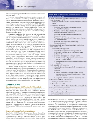
1953 and clearly distinguished the disorder from other acquired hemo- TABLE 54–1. Classification of Hemolytic Anemia as a
lytic syndromes.
In current usage, cold agglutinin disease pertains to patients with Result of Immune Injury
chronic AHA in which the autoantibody directly agglutinates human I. Warm-Autoantibody Type: Autoantibody Maximally Active at
RBCs at temperatures below body temperature, maximally at 0 to 5°C. Body Temperature (37°C)
Fixation of complement to a patient’s RBCs by cold agglutinins in vivo A. Primary or idiopathic warm autoimmune hemolytic anemia
occurs at higher temperatures but generally less than 37°C. Cold agglu- (AHA)
tinins typically are IgM, although occasionally they may be immu- B. Secondary warm AHA
noglobulins of other isotypes. The cold agglutinins in chronic cold 1. Associated with lymphoproliferative disorders
agglutinin disease generally are monoclonal. Most cold agglutinins have (e.g., Hodgkin lymphoma)
specificity for oligosaccharide antigens (I or i) of the RBC (see “Origin 2. Associated with the rheumatic disorders, particularly
of Cold Agglutinins” below). systemic lupus erythematosus (SLE)
Donath and Landsteiner first described the cold hemolysin that 3. Associated with certain nonlymphoid neoplasms
bears their name in 1904. The Donath-Landsteiner antibody is respon- (e.g., ovarian tumors)
sible for complement-mediated hemolysis in paroxysmal cold hemo- 4. Associated with certain chronic inflammatory diseases
globinuria, a rare form of AHA in adults. The disorder is characterized (e.g., ulcerative colitis)
by recurrent episodes of massive hemolysis following cold exposure. 10,11 5. Associated with ingestion of certain drugs
A related form of hemolytic anemia occurs much more commonly in (e.g., α-methyldopa)
children (or young adults) as an acute, self-limited hemolytic process II. Cold-Autoantibody Type: Autoantibody Optimally Active at
following several types of viral syndromes. 10–16 The disease was recog- Temperatures <37°C
nized during the latter half of the 19th century, when the disease was
more common because of its association with congenital or tertiary A. Mediated by cold agglutinins
syphilis. With the advent of effective therapy for syphilis, this cause of 1. Idiopathic (primary) chronic cold agglutinin dis-
paroxysmal cold hemoglobinuria has almost disappeared. Now, recur- ease (usually associated with clonal B-lymphocyte
rent paroxysmal cold hemoglobinuria occurs very rarely in a chronic proliferation)
idiopathic form. 10,11 An increasing proportion of Donath-Landsteiner 2. Secondary cold agglutinin hemolytic anemia
autoantibody-mediated hemolytic anemias occurs as a single post- a. Postinfectious (e.g., Mycoplasma pneumoniae or
viral episode in children, without recurrent attacks (paroxysms). The infectious mononucleosis)
prognosis for such cases is excellent. Thus, rather than paroxysmal cold b. Associated with malignant B-cell lymphoproliferative
hemoglobinuria, a proposed term for this latter entity is Donath-Land- disorder
steiner hemolytic anemia. 13,14 B. Mediated by cold hemolysins
The first example of drug-related immune blood cell destruction 1. Idiopathic (primary) paroxysmal cold hemoglobinuria
17
was Ackroyd’s description of sedormid purpura in 1949. In 1953, Snap- (very rare)
per and coworkers described a case of immune hemolysis and pancy- 2. Secondary
18
topenia in a patient treated with mephenytoin (Mesantoin). Hemolysis
19
ceased upon withdrawal of the drug. In 1956, Harris reported what a. Donath-Landsteiner hemolytic anemia, usually
associated with an acute viral syndrome in children
are now classic studies of a patient who developed immune hemolytic (relatively common)
anemia during a second course of stibophen administered for treatment
of schistosomiasis. Since then, many drugs have been implicated in the b. Congenital or tertiary syphilis in adults (very rare)
production of positive DATs and accelerated RBC destruction. III. Mixed Cold and Warm Autoantibodies
A. Primary or idiopathic mixed AHA
CLASSIFICATION B. Secondary mixed AHA
1. Associated with the rheumatic disorders, particularly SLE
Warm-Reactive versus Cold-Reactive Red Cell Antibody IV. Drug-Immune Hemolytic Anemia
AHA can be classified in two complementary ways (Table 54–1). The
majority of cases (80 to 90 percent in adults) are mediated by warm- A. Hapten or drug adsorption mechanism
reactive autoantibodies 10,11,20 or antibodies displaying optimal reactivity B. Ternary (immune) complex mechanism
with human RBCs at 37°C. A smaller proportion of cases is attribut- C. True autoantibody mechanism
able to cold-reactive autoantibodies exhibiting greater affinity for RBCs
at temperatures less than 37°C. The distinction is important, not only
because of differences in the pathophysiology of RBC injury but also disorder, the term secondary AHA is applied. Lymphocytic malignan-
in the therapeutic approaches required. An even smaller proportion of cies, particularly chronic lymphocytic leukemia (CLL) and lymphomas,
patients with AHA exhibit both cold-reactive and warm-reactive auto- account for approximately half of all secondary AHA cases and for the
antibodies, 21,22 which apparently recognize different antigens on the majority of AHA cases mediated by cold agglutinins. Systemic lupus
24
RBC membrane. RBC destruction is generally more severe in mixed erythematosus (SLE) and other autoimmune diseases account for a lesser
23
cases. but considerable proportion of secondary AHA cases. A large propor-
tion of patients with mixed cold and warm autoantibodies have SLE. 21,22
Absence or Presence of an Associated Disease Infectious mononucleosis and Mycoplasma pneumoniae occasionally
Classification of AHA based on the presence or absence of underlying are associated with cryopathic AHA. Despite the frequent occurrence of
diseases also is useful (see Table 54–1). When no recognizable underly- immune thrombocytopenia and positive DATs in patients infected with
ing disease is present, the AHA is termed primary or idiopathic. When HIV, AHA is relatively rare in these patients. 25–27 Table 54–1 lists other
AHA appears to be a manifestation or complication of an underlying associated diseases that are less-commonly reported. The etiologic and
Kaushansky_chapter 54_p0823-0846.indd 824 9/19/15 12:26 AM