
Page 884 - Williams Hematology ( PDFDrive )
P. 884
858 Part VI: The Erythrocyte Chapter 55: Alloimmune Hemolytic Disease of the Fetus and Newborn 859
including two preparations that may be administered intravenously. A CONCLUSIONS
131
300 mcg (1500 IU) dose of RhIg affords protection against a fetomater-
nal transfusion of 15 mL of Rh-positive RBCs or 30 mL of Rh-positive HDFN is a clinically significant problem that may potentially affect
WB. However, FMH in excess of 30 mL may occur in women without any pregnancy. Although a number of strategies are in place to prevent
predisposing risk factors. 42,43 The blood of all Rh-negative nonimmu- RhD HDFN, few options exist to prevent the development of non-D
nized women should be tested for FMH approximately 1 hour after HDFN. Researchers continue to investigate strategies to prevent pri-
delivery of an Rh-positive baby. 57,129 During the antenatal period, testing mary maternal RBC alloimmunization to RhD and to non-D antigens,
is indicated after 20 weeks’ gestation if clinical circumstances suggest as well as strategies to mitigate the dangers of existing maternal RBC
the possibility of excessive transplacental hemorrhage (e.g., abdominal alloantibodies. Over the past decade, the ability to identify pregnancies/
trauma or abruptio placentae). Screening for FMH can be performed fetuses “at risk” because of maternal RBC alloimmunization has signifi-
by the rosette test, which detects as little as 2.5 mL of WB. If the rosette cantly improved. In particular, the use of noninvasive investigations,
test result is positive, the number of fetal red cells in the maternal circu- such as evaluation of fetal RBC antigen zygosity by circulating cell free
lation is quantified more accurately by the Kleihauer-Betke test, which fetal DNA testing and the evaluation of fetal anemia by middle cerebral
is based on the resistance of fetal hemoglobin to acid elution, unlike artery Doppler ultrasonography, have advanced the care of fetuses at
132
adult hemoglobin (Fig. 55–5). False-positive results can be obtained risk for HDFN. Through the continued combined efforts of maternal–
in maternal conditions associated with increased fetal hemoglobin, fetal medicine specialists, hematologists, transfusion medicine phy-
such as hereditary persistence of fetal hemoglobin, sickle cell disease, or sicians, radiologists, neonatologists, and researchers, in combination
sickle cell trait. Flow cytometric methods are used in some laboratories with advancements in basic science research involving alloimmuniza-
for both screening and quantification of fetal red cells. tion, the care for infants at risk for HDFN, and their mothers, will likely
RhIg should be administered as soon as possible, within 72 hours continue to improve in the years to come.
of delivery of an Rh-positive baby. RhIg is thought to be ineffective
once alloimmunization to RhD antigen has occurred. RhIg is also indi-
cated following pregnancy termination, miscarriage, amniocentesis, REFERENCES
chorionic villus sampling, or other manipulation during pregnancy. A
smaller 50-mcg dose is adequate if pregnancy is terminated at less than 1. Diamond L, Blackfan K, Baty J: Erythroblastosis fetalis and its association with univer-
sal edema of the fetus, icterus gravis neonatorum and anemia of the newborn. J Pediatr
129
12 weeks’ gestation. If therapeutic or spontaneous abortion occurs 1932(1):269.
after the first trimester, the standard 300-mcg dose is recommended. 2. Darrow R: Icterus gravis (erythroblastosis neonatorum, examination of etiologic con-
129
siderations). Arch Pathol 1938(25):378.
If a woman was exposed to more than 30 mL of D-positive blood, the 3. Levine P, Katzin E, Burnham L: Isoimmunization in pregnancy: Its possible bearing on
dose of RhIg should be calculated to cover the volume of D-positive the etiology of erythroblastosis fetalis. JAMA 116(9):825, 1941.
cells to prevent immunization (20 mcg of RhIg for 1 mL of D+ RBCs 4. Diamond LK, Allen FH Jr, Thomas WO Jr: Erythroblastosis fetalis. VII. Treatment with
or 2 mL of WB). 57,131,133 Despite the efficacy of RhIg, RhD alloimmuni- exchange transfusion. N Engl J Med 244(2):39, 1951.
zation occurs despite recommended prophylaxis in 0.1 percent of preg- 5. Liley AW: The use of amniocentesis and fetal transfusion in erythroblastosis fetalis.
Pediatrics 35:836, 1965.
nancies, but many cases of preventable Rh alloimmunization continue 6. Bowman J: Thirty-five years of Rh prophylaxis. Transfusion 43(12):1661, 2003.
to occur because of failure to seek medical care or because of failure to 7. Poole J, Daniels G: Blood group antibodies and their significance in transfusion medi-
cine. Transfus Med Rev 21(1):58, 2007.
implement immunoprophylaxis protocols. 8. Geifman-Holtzman O, et al: Female alloimmunization with antibodies known to cause
hemolytic disease. Obstet Gynecol 89(2):272, 1997.
9. Gottvall T, Filbey D: Alloimmunization in pregnancy during the years 1992-2005 in the
central west region of Sweden. Acta Obstet Gynecol Scand 87(8):843, 2008.
10. Koelewijn JM, Vrijkotte TG, van der Schoot CE, et al: Effect of screening for red cell
antibodies, other than anti-D, to detect hemolytic disease of the fetus and newborn: A
population study in the Netherlands. Transfusion 48(5):941, 2008.
11. Lee CK, Ma ES, Tang M, et al: Prevalence and specificity of clinically significant red cell
alloantibodies in Chinese women during pregnancy—A review of cases from 1997 to
2001. Transfus Med 13(4):227, 2003.
12. Zipursky A, Paul VK: The global burden of Rh disease. Arch Dis Child Fetal Neonatal Ed
96(2):F84, 2011.
13. Moise KJ: Fetal anemia due to non-Rhesus-D red-cell alloimmunization. Semin Fetal
Neonatal Med 13(4):207, 2008.
14. Garratty G, Glynn SA, McEntire R: ABO and Rh(D) phenotype frequencies of different
racial/ethnic groups in the United States. Transfusion 44(5):703, 2004.
15. Joseph KS: Controlling Rh haemolytic disease of the newborn in India. Br J Obstet
Gynaecol 98(4):369, 1991.
16. Mak KH, Yan KF, Cheng SS, Yuen MY: Rh phenotypes of Chinese blood donors in
Hong Kong, with special reference to weak D antigens. Transfusion 33(4):348, 1993.
17. Ziprin JH, Payne E, Hamidi L, et al: ABO incompatibility due to immunoglobulin G
anti-B antibodies presenting with severe fetal anaemia. Transfus Med 15(1):57, 2005.
18. Thilaganathan B, Salvesen DR, Abbas A, et al: Fetal plasma erythropoietin concentra-
tion in red blood cell-isoimmunized pregnancies. Am J Obstet Gynecol 167(5):1292,
1992.
19. Koenig JM, Christensen RD: Neutropenia and thrombocytopenia in infants with Rh
hemolytic disease. J Pediatr 114(4 Pt 1):625, 1989.
20. Hayde M, Widness JA, Pollak A, et al: Rhesus isoimmunization: increased hemolysis
during early infancy. Pediatr Res 41(5):716, 1997.
21. al-Alaiyan S, al Omran A: Late hyporegenerative anemia in neonates with rhesus hemo-
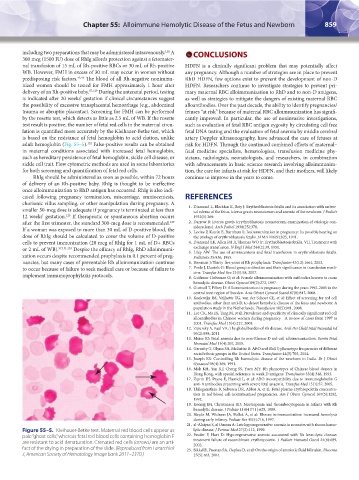
Figure 55–5. Kleihauer-Betke test. Maternal red blood cells appear as lytic disease. J Perinat Med 27(2):112, 1999.
pale “ghost cells,” whereas fetal red blood cells containing hemoglobin F 22. Pessler F, Hart D: Hyporegenerative anemia associated with Rh hemolytic disease:
are resistant to acid denaturation. Crenated red cells (arrows) are an arti- treatment failure of recombinant erythropoietin. J Pediatr Hematol Oncol 24(8):689,
2002.
fact of the drying in preparation of the slide. (Reproduced from Lazarchick 23. Sikkel E, Pasman SA, Oepkes D, et al: On the origin of amniotic fluid bilirubin. Placenta
J, American Society of Hematology Image bank 2011–2370.) 25(5):463, 2004.
Kaushansky_chapter 55_p0847-0862.indd 859 9/18/15 11:53 PM