
Page 973 - Williams Hematology ( PDFDrive )
P. 973
948 Part VII: Neutrophils, Eosinophils, Basophils, and Mast Cells Chapter 62: Eosinophils and Related Disorders 949
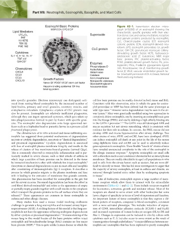
Figure 62–1. Transmission electron micro-
graph (×10,000) of an eosinophil showing the
characteristic specific granules with their elec-
tron dense core and various mediators, receptors
and granule proteins produced by eosinophils.
CLC, Charcot-Leyden crystal; ECP, eosinophil
cationic protein; EDN, eosinophil-derived neu-
rotoxin; EPO, eosinophil peroxidase; GF, growth
factor; GM-CSF, granulocyte-monocyte colony-
stimulating growth factor; HETE, hydroxyeico-
satetraenoic acid; LT, leukotriene; MBP, major
basic protein; PAF, platelet-activating factor;
PDGF, platelet-derived growth factor; PG, pros-
taglandin; PSGL, P-selectin glycoprotein ligand;
TBX, thromboxane; TGF-β, transforming growth
factor-β; VEGF, vascular endothelial growth fac-
tor. (Used with permission of Dr. A. Dewar, National
Heart and Lung Institute.)
into specific granules. Electron microscopy can distinguish acti- cell free basic proteins can be readily detected in both tissue and BAL.
vated from resting blood eosinophils by the increased number of Consistent with this observation, mice in which the gene for eosino-
lipid bodies, primary and small granules, secretory vesicles, and phil peroxidase or MBP has been deleted had the same phenotype as
endoplasmic reticulum. Cytoplasmic crystals of CLC protein may wild-type mice. However some degranulation may be seen in the air-
13
also be present. Eosinophils are relatively inefficient phagocytes, way lumen. Mice were genetically modified using two approaches to
14
although they can ingest opsonized zymosan, which gets taken up completely delete eosinophils, one by inserting an eosinophil toxic gene
into phagolysosomes formed in part by fusion with specific gran- into the lineage (PHIL) and one by deleting a high-affinity binding site
ules. The eosinophil also degranulates onto large opsonized sur- in the GATA-1 promoter. 15,16 The GATA mice still developed AHR and
faces such as a Sephadex bead or parasitic larvae in a process called mucus secretion but not airway remodeling, which is consistent with
frustrated phagocytosis. evidence for their role in asthma. In contrast, the PHIL mouse did not
The ultrastructure of in vitro activated and tissue-infiltrating eos- develop AHR and mucus hypersecretion after airway challenge. Two
inophils has suggested three potential mechanisms of degranulation: other strains of mice, iPHIL and eoCRE, have been developed. iPHIL
necrosis or cytolytic degranulation, exocytosis or “classical degranulation,” can induce eosinophil cell death at any point in the life of the mouse
and piecemeal degranulation. Cytolytic degranulation is associated using diphtheria toxin and eoCRE can be used to selectively induce
7
with loss of eosinophil plasma membrane integrity and results in the gene expression in eosinophils. These flexible “knock-in” strains of mice
release of clusters of free membrane-bound granules (termed Cfegs). have revealed unexpected complexity in the role of the eosinophil in
This is commonly observed in eosinophilic inflammation and is par- the allergic immune response. Apoptotic eosinophils are small cells
17
ticularly marked in severe disease, such as fatal attacks of asthma in with a shrunken nucleus and condensed chromatin but an intact plasma
which large quantities of basic proteins can be detected in the tissue membrane. They are readily identifiable in aged cell populations in vitro
by immunohistochemistry often with relatively few intact eosinophils. and in cells from the airway lumen such as sputum, but are more dif-
8
Exocytosis or classical degranulation occurs in mast cells and basophils ficult to identify in tissue. This has led some investigators to argue that
after crosslinking of immunoglobulin (Ig) E receptors. It describes a the majority of airway eosinophils, at least in asthma and rhinitis, are
process by which granules migrate to the plasma membrane and fuse removed through luminal entry rather than by undergoing apoptosis
with it leading to the extrusion of membrane free granule contents. in tissue. 7
This phenomenon has been described for eosinophils in the gut, but Like all leukocytes, eosinophils express a large number of mem-
not the airway mucosa. Piecemeal degranulation has been described in brane receptors which allow them to interact with the extracellular
cord blood derived eosinophils and refers to the appearance of empty environment (Tables 62–1 and 62–2). These include receptors required
9
or partially empty granules together with small vesicles in the cytoplasm for locomotion, activation, growth and mediator release. Most of the
which transport the granule proteins to the cell surface where they are receptors are shared to some extent with other leukocytes but some
released. These appearances are common in tissue eosinophils in have a degree of specificity in terms of level of expression and function.
10
asthma and other allergic diseases. An important feature of tissue eosinophils is that they express a dif-
Many studies have used a mouse model involving ovalbumin ferent pattern of receptors, compared to blood eosinophils, consistent
challenge to generate a lung eosinophilia and increased airway hyper- with a more activated phenotype. This includes induction of expres-
responsiveness (AHR). A striking feature of this model is that lung eos- sion of CD69, intercellular adhesion molecule (ICAM)-1 and FcγR1
inophils do not have the appearance of having undergone degranulation and increased expression of human leukocyte antigen (HLA)-DR and
by either cytolysis or piecemeal degranulation. Immunostaining of the Mac-1. Changes in expression can be induced in vitro by culture with
11
mouse lung in this model locates all the basic proteins within intact cytokines such as IL-5, but also occur to some extent as the result of
eosinophils and bronchoalveolar lavage (BAL) contains no free major transmigration through endothelium. A major difference between eos-
18
basic protein (MBP). This is quite unlike human disease in which the inophils and neutrophils that has been exploited to purify eosinophils
12
Kaushansky_chapter 62_p0947-0964.indd 948 9/21/15 10:56 AM