
Page 974 - Williams Hematology ( PDFDrive )
P. 974
948 Part VII: Neutrophils, Eosinophils, Basophils, and Mast Cells Chapter 62: Eosinophils and Related Disorders 949
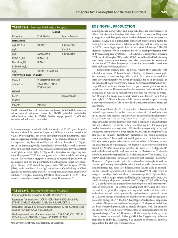
TABLE 62–1. Eosinophil Adhesion Receptors EOSINOPHIL PRODUCTION
Ligand Eosinophils are nondividing, end-stage cells that, like other leukocytes,
differentiate from the hematopoietic stem cell in the marrow. They share
Receptor Endothelial Matrix Protein a progenitor with basophils before further differentiation separates the
INTEGRINS lineages. GATA-1 is a particularly important transcription factor for
α β (VLA-4) VCAM-1 Fibronectin eosinophil development with deletion of the high-affinity binding site
4 1
in GATA-1 resulting in specific loss of the eosinophil lineage. The F/P
29
α β Laminin
4 6 receptor mutation which is responsible for a myeloproliferative form
α β MAdCAM-1 Fibronectin of hypereosinophilic syndrome (HES)/chronic eosinophilic leukemia
4 7
LFA-1 (α β ) ICAM-1-3 (CEL) works through cEBPα and GATA-2, as well as GATA-1, showing
Λ 2 that these transcription factors are also important in eosinophil
Mac-1 (α β ) ICAM-1 development. Eosinophilopoiesis requires the combined expression of
30
M 2
P150,95 (α β ) MBP and eosinophil peroxidase. 31
x 2
α β VCAM-1 (ICAM-3?) Eosinophils migrate into the blood, where they circulate with
d 2 a half-life of about 18 hours before entering the tissues. Eosinophils
SELECTINS AND LIGANDS are primarily tissue-dwelling cells, and it has been estimated that
PSGL-1 P-selectin(E-selectin) there are approximately 100 tissue eosinophils for each eosinophil in
the blood, although relatively few studies have been performed on eos-
l-Selectin Gly-CAM-1, CD34,
Podocalyxin inophil kinetics and even fewer have compared eosinophil turnover in
health and disease. However, studies demonstrate that eosinophils can
OTHER be tracked in vivo using radiolabeling and that the kinetics of migra-
CD44 Hyaluronate tion through the lung, spleen, and marrow are distinct from that of
ICAM-3 neutrophils. 32,33 Normal human adult marrow contains approximately
3 percent eosinophils of which one-third are mature and two-thirds are
PECAM PECAM precursors.
ICAM, intercellular cell adhesion molecule; MAdCAM-1, mucosal Eosinophilia is often T-cell dependent. Characterization of T-cell–
addressin cell adhesion molecule; PECAM: platelet endothelial derived supernatants led to the characterization of IL-5 and awareness
34
cell adhesion molecule; PSGL-1, P-selectin glycoprotein 1; VCAM-1, of the pivotal role that this cytokine plays in eosinophil development.
vascular cell adhesion molecule. IL-3 and GM-CSF are also important in eosinophil development. The
three cytokines bind to receptors that share a common β chain but have
distinct α chains. IL-5 seems to be a rate-limiting step for eosinophil
by immunomagnetic selectin is the expression of CD16 by neutrophils production in that administration of IL-5 either exogenously or through
35
but not eosinophils. Another important difference is the expression of transgenic manipulation in mice results in a marked eosinophilia and
VLA-4 by eosinophils, but not to any great extent in neutrophils. Sialic anti–IL-5 in humans dramatically diminishes the blood eosinophil
36
acid-recognizing animal lectin (Siglec) 8 has been identified as a recep- count in asthma. Increased eosinophilopoiesis as a result of increased
tor expressed only by eosinophils, mast cells, and basophils. 19–21 Siglecs IL-5 synthesis appears to be a feature of a number of diseases, includ-
are of the immunoglobulin superfamily. Eosinophils, as well as mono- ing parasitic and allergic diseases. For example, pulmonary eosinophilia
37
22
cytes and a subset of dendritic cells, also express Siglec 10. In contrast, caused by Necator americanus infection in mice is IL-5–dependent
23
neutrophils express Siglec 9. Siglec 8 is important in triggering eos- and both the eosinophilia and host defense to filariasis and Trichinella
38
inophil apoptosis. 24,25 Epidermal growth factor-like module containing spiralis is markedly impaired in IL-5–deficient mice. In asthma, IL-5
39
mucin-like hormone receptor 1 (EMR1) is expressed exclusively on mRNA can be detected in increased amounts in the airways in asthma.
eosinophils and has the potential to be a therapeutic target for eosino- However, IL-5 gene-deleted mice have a baseline eosinophilia and can
philic disorders. Eosinophils express both CD48 and its ligand CD244 develop pulmonary eosinophilia after infection with paramyxovirus,
26
(2B4), both members of the IgG superfamily. Crosslinking of CD48 demonstrating that other late differentiation factors such as chemok-
27
causes eosinophil degranulation. Eosinophils also express a number of ine (C-C motif) ligand (CCL)-3 may be involved. 40,41 It is therefore an
inhibitory receptors including CMRF35-like molecule-1 (CLM-1) that accepted paradigm that a blood and tissue eosinophilia in IgE-mediated
negatively regulate eotaxin-induced eosinophil responses. 28 diseases, such as atopic asthma and helminthic parasite infections, are a
result of antigen-dependent activation of T-helper (Th)-2 cells leading
to IL-5 production and increased eosinophilopoiesis and tissue recruit-
ment of eosinophils. The control of development of Th2 and Th1 cells is
TABLE 62–2. Eosinophil Adhesion Receptors beyond the scope of this chapter, but may relate to the cytokine milieu
at the time of sensitization, genetically regulated transcriptional control
Immunoglobulin receptors: Fcγ R11 (CD32); Fcα R;
of IL-4, or the route of sensitization and the way in which the antigen is
Receptors for mediators: CCR3*; CCR1; PAF-R; LTC4/D4/E4-R; presented (Chap. 76). 42,43 The HLA haplotype of individuals responsive
LTB4-R; C5aR; C3aR; IL-5R*; IL-3R; IL-4R; IL-13R; CRTh2 to certain allergens has also been investigated. A degree of restriction
Receptors induced by cytokine stimulation: Fcγ RIII (CD16); Fcγ R1 has been observed, particularly to simple allergens, with, for example,
(CD69); HLA-DR; ICAM-1; CD25; CD4 the phenotype DR2.2 being overrepresented in individuals atopic to the
Well-expressed miscellaneous receptors: CD9; CD45; CR1; CD154 ragweed allergen Amb a V. However, with the majority of allergens, no
(CD40 ligand); CD95 (Fas); Siglec 8*; ERM1*; CLM-1 clear pattern has emerged. Although HLA haplotypes may influence
responses to individual allergens, it is unlikely to provide a universal
*Relatively selectively expressed by eosinophils. explanation for Th2-type responsiveness.
Kaushansky_chapter 62_p0947-0964.indd 949 9/21/15 10:56 AM