
Page 976 - Williams Hematology ( PDFDrive )
P. 976
950 Part VII: Neutrophils, Eosinophils, Basophils, and Mast Cells Chapter 62: Eosinophils and Related Disorders 951
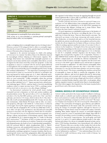
TABLE 62–3. Eosinophil Chemokine Receptors and also regulated by the balance between the signaling through two paired
immunoglobulin-like receptors, PIR-A and PIR-B, with PIR-B counter-
Their Ligands
acting the proapoptotic effect of PIR-A. 81
Receptor Chemokine Another potential mechanism involved in eosinophil tissue accu-
CCR1* CCL3 (Mip-1a); CCL5 (RANTES) mulation is in situ differentiation from eosinophil precursors. Eosin-
ophil precursors can be identified in an IL-5Rα CD34 population in
+
+
CCR3 CCL11 (eotaxin 1); CCL24 (eotaxin 2); CCL26 blood, increased after allergen challenge and in atopic disease. These
(eotaxin 3); CCL7, -8, -13 (MCP2–4); CCL5
cells have also been found in asthmatic airways. 82
CXCR1, -2 CXCL8 (IL-8 †) Of equal importance as endothelial interactions to the kinetics of
eosinophil migration are the factors controlling the fate of the eosin-
*Only expressed on eosinophils from some donors. ophil once it enters the tissue. There are three possible outcomes. The
† Only active on in vivo–activated or cytokine-primed eosinophils eosinophil can remain in the tissue interacting with matrix proteins,
(may be indirect effect via neutrophils).
other leukocytes, or structural cells such as, in the bronchial mucosa,
the epithelium, airway smooth muscle, mucus glands, and nerves; alter-
natively, the cell can migrate into the lumen of the gut or airway where it
to play overlapping roles in eosinophil migration into the lung in mice. is likely to undergo apoptosis and be removed; or it can return to the cir-
69
However, a potent CCR3 antagonist had no effect on eosinophil migra- culation via the lymphatics. The length of time that eosinophils remain
tion into the airways in human asthma, questioning the physiologic rel- in tissue before migrating into the lumen is unclear as there are virtually
evance of the mouse data in human disease. 70 no studies of the kinetics of eosinophil migration in vivo in humans.
Apoptosis is the universal mechanism by which cells undergo An anti–IL-5 completely inhibited migration into the lumen, which
cell senescence in a manner that allows them to be efficiently removed suggests that transepithelial migration is IL-5–dependent. However, it
by macrophages without inducing an inflammatory response. Mor- only inhibited tissue numbers by at best 50 percent, emphasizing that
phologic observations have indicated that eosinophil apoptosis is an different compartments are controlled by different mechanisms. In
73
unusual event in tissue and that most eosinophils either die by cytolysis the mouse model of asthma, eosinophil migration into the lumen does
or migrate into the lumen where they do become apoptotic. A slow rate not occur in the MMP-2 gene-deleted mouse, and the lack of migration
7
of apoptosis in tissues is consistent with the survival signals delivered causes the mouse to asphyxiate. As with senescent neutrophils, when
83
to eosinophils by the extracellular matrix as part of normal homeosta- tissue eosinophils become senescent they start to alter their receptor
sis as well as increased production of eosinophil growth factors during phenotype in a way that inhibits tissue retention and promotes migra-
84
Th2-mediated inflammation. 71,72 The importance of prolonged survival tion into the lumen. The factors controlling the retention and survival
of eosinophils in tissue as a mechanism for selective accumulation has of eosinophils in tissue are likely to involve the integration of che-
been emphasized by studies using anti–IL-5, which effectively inhib- moattractant, adhesive, and survival signals delivered by interactions
its blood and sputum eosinophil numbers but has a much-less-marked with matrix proteins and structural cells. Studies modeling eosinophil
effect on tissue eosinophils. Unlike neutrophils, where they prolong migration in a tissue context using collagen gels have shown a different
73
survival, glucocorticoids directly enhance the rate of eosinophil apop- pattern to standard Boyden chamber assays with a much greater, albeit
85
tosis, an effect inhibited by IL-5. Tumor necrosis factor-related apop- random, migratory response to growth factors than to chemoattractants.
74
tosis-inducing ligand (TRAIL), another family of survival modulating This observation suggests that migration into the lumen requires both a
mediators related to tumor necrosis factor (TNF)-α, prolongs eosino- growth factor and a chemotactic stimulus.
phil survival, both in vitro and ex vivo, after allergen challenge. 75
The biochemical mechanism by which growth factors mediate
eosinophil survival is dependent on both new protein synthesis and ANIMAL MODELS OF EOSINOPHILIC DISEASE
phosphorylation events. The survival effects of IL-5 are dependent on Animal models, particularly the mouse model of ovalbumin challenge,
activation of the Ras-Raf-MEC pathway and the Jak-2 Stat 1 and Stat 5 which results in a selective and marked pulmonary eosinophilia, have
pathways, and involve LYN kinase, which binds to the IL-5Rα chain. been used extensively to analyze the molecular basis of eosinophil traf-
76
The roles of p38 and phosphatidylinositide (PI) 3 kinase are less clear, ficking and the pathologic consequences of this movement. The major-
86
and wortmannin, which blocks PI3 kinase, had no effect on eosino- ity of studies have focused on the role of eosinophilic inflammation in
phil apoptosis, although it did inhibit IL-5 enhancement of adhesion asthma. The combination of transgenic, gene-deletion, and antibody-
87
to fibrinogen. Eosinophils express significant amounts of the proapop- based manipulations in the mouse make this a powerful tool for ana-
totic BAX and the antiapoptotic BCL-xl, but very little Bad or BCL-2. lyzing the biology of eosinophil migration, although the relevance of
As in other cell types, both spontaneous and FAS-induced eosinophil the findings to human disease should be treated with caution. Generally
apoptosis is associated with the migration of BAX into the mitochon- speaking, these studies support the concept of eosinophil migration
dria. This event led to loss of mitochondrial membrane potential, cyto- as being caused by a series of interlinked and obligate steps with IL-5
chrome c release, and activation of downstream caspases. These events necessary for providing a pool of circulating eosinophils, priming eos-
are all inhibited by IL-5, demonstrating that IL-5 works by blocking inophils for chemotactic responsiveness, and prolonging eosinophil
BAX translocation. 77,78 Inhibition of BAX activation prevents eosino- survival. IL-4 and IL-13 control adhesion-related events in the endo-
phil apoptosis even in the absence of cytokine. Treatment of eosinophils thelium and enhance the release of eosinophil chemoattractants, partic-
with dexamethasone also leads to loss of mitochondrial permeability. ularly CCR3-binding chemokines from mesenchymal cells within the
79
GM-CSF–activated ERK1/2, which phosphorylates BAX at Thr167, airway. However, there are a number of other studies that look at other
58
facilitates interaction with peptidylprolyl isomerase (PIN1). If interac- aspects of the immune response and as a result challenge this neat con-
tion with PIN1 is prevented, BAX is activated and translocated to the cept, in particular showing potential roles for innate immunity as well
mitochondria, resulting in apoptosis. It appears, therefore, that the eos- as other inflammatory mediators. 88–90 Our understanding of the role of
inophil growth factors exert their antiapoptotic effects through foster- eosinophils in health and disease, using mouse models of disease, has
ing the PIN1–BAX interaction. IL-5–mediated eosinophil survival is been summarized. 17
80
Kaushansky_chapter 62_p0947-0964.indd 951 9/21/15 10:56 AM