
Page 982 - Williams Hematology ( PDFDrive )
P. 982
956 Part VII: Neutrophils, Eosinophils, Basophils, and Mast Cells Chapter 62: Eosinophils and Related Disorders 957
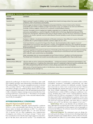
TABLE 62–5. Helminthic Causes of an Eosinophilia
Parasite Comment
NEMATODES
Ascariasis Higher eosinophil counts in children. Larvae migrate from intestine to lungs where they cause Loeffler
syndrome, a form of pulmonary eosinophilia.
Toxocara canis Infective eggs are present in feces of puppies and pregnant bitches. Larvae in hosts such as chicken.
Eosinophilia seen mainly in children younger than 9 years of age. Can migrate to eye and cause blindness.
Serologic evidence suggests infection not uncommon in industrialized countries.
Filariasis Common. Invariably result in marked eosinophilia, especially Loa Loa infection. Filariasis causes tropical
pulmonary eosinophilia as a result of migration of adult worms to the lung, elephantiasis because of the
involvement of lymphatics (Wuchereria bancrofti and Brugia malayi), and river blindness (Onchocerca volvulus).
Treatment can result in systemic reaction called Mazzotti reaction, possibly a result of massive eosinophil
degranulation.
Ancylostomiasis Hookworm infection. Ancylostoma duodenale and Necator americanus. One of the main causes of eosinophilia
9
in patients returning from tropical countries. Counts in region of 2.0 × 10 /L.
Strongyloidiasis Subclinical infection can persist for longer than 20 years. Stool examinations often negative. Cause of
eosinophilia in ex-servicemen who spent time in tropics. If strongyloides infection is not considered and these
patients are given steroids for suspected hypereosinophilic syndrome or as trial of therapy, they can develop
disseminated disease.
Trichinosis Caused by ingestion of encysted muscle larvae of Trichinella spiralis. Most prominent eosinophilia seen during
early stages of infection when larvae migrating into striated muscle via the blood. Fatal cases reported of
which only 20% were noted to have an eosinophilia.
Others Other nematodes that can cause eosinophilia include Trichuris trichiura, Capillaria, and Gnathostomiasis.
The thread worm Enterobius vermicularis occasionally causes an eosinophilia when they invade tissues.
TREMATODES
Schistosomiasis (Bilharzia) Infection with one of the Schistosoma (blood flukes)— Schistosoma mansoni, Schistosoma haematobium, and
Schistosoma japonicum—is perhaps the commonest cause of a moderate to high eosinophilia worldwide, with
200 million people being infected. Infection is nearly always associated with an eosinophilia.
Fascioliasis Adult worms of Fasciola hepatica reside in the bile ducts, where they are associated with abnormal liver
function tests and an eosinophilia.
CESTODES
Echinococcus Eosinophilia occurs in 25–50% of patients with hydatid disease.
infection by producing IL-10 which led to a reduction in nitric oxide classification of HES is unsatisfactory as conditions such as EGPA,
(NO) synthase expression and protected the intracellular larvae from eosinophilic pneumonia, eosinophilic gastroenteritis, and other
NO-mediated killing. In contrast, eosinophils were protective for host more organ-specific conditions are arbitrarily excluded. Attempts
148
with a secondary infection. The mechanism of eosinophilia in para- have been made to create a more comprehensive and less arbitrary
149
sitic disease is thought to be similar to allergic disease, with a Th2-type system although this requires more data on specific etiologies. 155,156
response mediated by both adaptive Th2 cells and innate lymphoid type The World Health Organization (WHO) takes the view that all
2 cells to helminthic antigens resulting in increased production of eos- myeloproliferative variants of HES are malignant conditions. It pre-
inophil growth factors, in particular IL-5. Recruitment of eosinophils viously classified them under chronic myeloproliferative diseases
150
to the site of parasite infection may involve nematode induced produc- (CMPDs) with CEL/HES as a subcategory. In WHO’s 2008 revision
tion of LTB by eosinophils. 151 they have called CMPD myeloproliferative neoplasms. Where there
4
is no defined genotypic abnormality the condition has been termed
CEL-NOS (not otherwise specified), and where there is a defined
HYPEREOSINOPHILIC SYNDROMES mutation such as F/P they fall into a new category of their own. 157
Idiopathic Hypereosinophilic Syndrome Epidemiology HES is a rare disorder with an estimated preva-
Definition and History Idiopathic hypereosinophilic syndrome lence (although there are limited data on this), in the region of one in
(iHES) is a rare and potentially fatal disorder first described as a 50,000. It occurs sporadically and there appears to be no geographic or
152
distinct entity by Hardy and Anderson in 1968. It is defined as environmental influences. For unknown reasons the myeloproliferative
9
a persistent eosinophilia of greater than 1.5 × 10 cells/L for more form has a strong male bias.
than 6 months with no other explanation after comprehensive Differential Diagnosis The commonest causes are allergic dis-
153
investigation and evidence of end-organ damage. Patient series ease and in particular allergy to thermotolerant colonizing fungi such as
have consistently shown that the major target organs for tissue dam- Aspergillus fumigatus and Candida albicans, chronic infection with hel-
age are the skin, heart, and nervous system. The definition and mintic parasites (see Table 62–5), severe asthma, chronic eosinophilic
154
Kaushansky_chapter 62_p0947-0964.indd 957 9/21/15 10:56 AM