
Page 981 - Williams Hematology ( PDFDrive )
P. 981
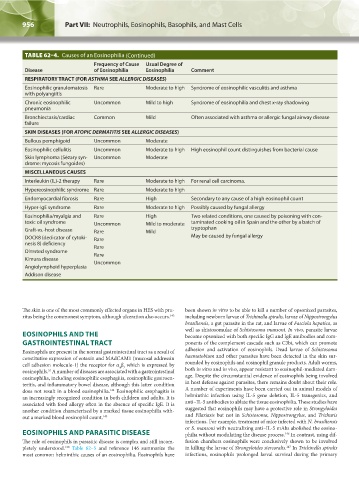
956            Part VII:  Neutrophils, Eosinophils, Basophils, and Mast Cells                                                                                            Chapter 62:  Eosinophils and Related Disorders            957


                TABLE 62–4.  Causes of an Eosinophilia   (Continued)
                                       Frequency of Cause   Usual Degree of
                Disease                of Eosinophilia   Eosinophilia   Comment
                RESPIRATORY TRACT (FOR ASTHMA SEE ALLERGIC DISEASES)
                Eosinophilic granulomatosis  Rare        Moderate to high  Syndrome of eosinophilic vasculitis and asthma
                with polyangiitis
                Chronic eosinophilic   Uncommon          Mild to high   Syndrome of eosinophilia and chest x-ray shadowing
                pneumonia
                Bronchiectasis/cardiac   Common          Mild           Often associated with asthma or allergic fungal airway disease
                failure
                SKIN DISEASES (FOR ATOPIC DERMATITIS SEE ALLERGIC DISEASES)
                Bullous pemphigoid     Uncommon          Moderate
                Eosinophilic cellulitis  Uncommon        Moderate to high  High eosinophil count distinguishes from bacterial cause
                Skin lymphoma (Sézary syn-  Uncommon     Moderate
                drome: mycosis fungoides)
                MISCELLANEOUS CAUSES
                Interleukin (IL)-2 therapy  Rare         Moderate to high  For renal cell carcinoma.
                Hypereosinophilic syndrome Rare          Moderate to high
                Endomyocardial fibrosis  Rare            High           Secondary to any cause of a high eosinophil count
                Hyper-IgE syndrome     Rare              Moderate to high  Possibly caused by fungal allergy
                Eosinophilia/myalgia and   Rare          High           Two related conditions, one caused by poisoning with con-
                toxic oil syndrome     Uncommon          Mild to moderate  taminated cooking oil in Spain and the other by a batch of
                Graft-vs.-host disease  Rare             Mild           tryptophan
                DOCK8 (dedicator of cytoki-  Rare                       May be caused by fungal allergy
                nesis 8) deficiency    Rare
                Olmsted syndrome       Rare
                Kimura disease         Uncommon
                Angiolymphoid hyperplasia
                Addison disease


               The skin is one of the most commonly effected organs in HES with pru-  been shown in vitro to be able to kill a number of opsonized parasites,
               ritus being the commonest symptom, although ulceration also occurs. 143  including newborn larvae of Trichinella spiralis, larvae of Nippostrongylus
                                                                      brasiliensis, a gut parasite in the rat, and larvae of Fasciola hepatica, as
                                                                      well as shistosomulae of Schistosoma mansoni. In vivo, parasite larvae
               EOSINOPHILS AND THE                                    become opsonized with both specific IgG and IgE antibodies and com-
               GASTROINTESTINAL TRACT                                 ponents of the complement cascade such as C3bi, which can promote
                                                                      adhesion and activation of eosinophils. Dead  larvae of  Schistosoma
               Eosinophils are present in the normal gastrointestinal tract as a result of
               constitutive expression of eotaxin and MAdCAM1 (mucosal addressin   haematobium and other parasites have been detected in the skin sur-
               cell adhesion molecule-1) the receptor for α β  which is expressed by   rounded by eosinophils and eosinophil granule products. Adult worms,
                                                4 7
               eosinophils.  A number of diseases are associated with a gastrointestinal   both in vitro and in vivo, appear resistant to eosinophil-mediated dam-
                        55
               eosinophilia, including eosinophilic esophagitis, eosinophilic gastroen-  age. Despite the circumstantial evidence of eosinophils being involved
               teritis, and inflammatory bowel disease, although this latter condition   in host defense against parasites, there remains doubt about their role.
               does not result in a blood eosinophilia.  Eosinophilic esophagitis is   A number of experiments have been carried out in animal models of
                                             144
               an increasingly recognized condition in both children and adults. It is   helminthic infection using IL-5 gene deletion,  IL-5 transgenics,  and
               associated with food allergy often in the absence of specific IgE. It is   anti–IL-5 antibodies to ablate the tissue eosinophilia. These studies have
               another condition characterized by a marked tissue eosinophilia with-  suggested that eosinophils may have a protective role in Strongyloides
               out a marked blood eosinophil count. 145               and  Filariasis but not in  Schistosoma, Nippostrongylus, and  Trichuris
                                                                      infections. For example, treatment of mice infected with N. brasiliensis
                                                                      or S. mansoni with neutralizing anti–IL-5 mAbs abolished the eosino-
               EOSINOPHILS AND PARASITIC DISEASE                      philia without modulating the disease process.  In contrast, using dif-
                                                                                                       146
               The role of eosinophils in parasitic disease is complex and still incom-  fusion chambers eosinophils were conclusively shown to be involved
               pletely understood.   Table 62–5 and reference 146 summarize the   in killing the larvae of Strongyloides stercoralis.  In Trichinella spiralis
                                                                                                        147
                              130
               most common helminthic causes of an eosinophilia. Eosinophils have   infections, eosinophils prolonged larval survival during the primary


          Kaushansky_chapter 62_p0947-0964.indd   956                                                                   9/21/15   10:56 AM