
Page 911 - Clinical Immunology_ Principles and Practice ( PDFDrive )
P. 911
880 PARt SEVEN Organ-Specific Inflammatory Disease
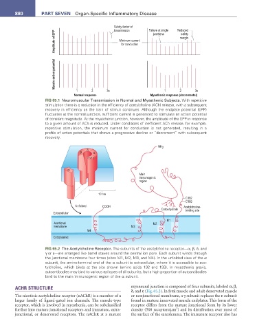
Safety factor of Failure at single Reduced
transmission
Amplitude of EPP Minimum current margin
junctions
safety
for conduction
Muscle action potential
1 2 3s 1 2 3s
Normal response Myasthenic response (decremental)
FIG 65.1 Neuromuscular Transmission in Normal and Myasthenic Subjects. With repetitive
stimulation there is a reduction in the efficiency of acetylcholine (ACh) release, with a subsequent
recovery in efficiency as the train of stimuli continues. Although the endplate potential (EPP)
fluctuates at the normal junction, sufficient current is generated to stimulate an action potential
of constant magnitude. At the myasthenic junction, however, the amplitude of the EPP in response
to a given amount of ACh is reduced. Under conditions of inefficient ACh release, for example,
repetitive stimulation, the minimum current for conduction is not generated, resulting in a
profile of action potentials that shows a progressive decline or “decrement” with subsequent
recovery.
NH 2
δ
β α
ε or γ
α
Main
immunogenic
region
10 nm
C192
C193
Unfolded COOH Acetylcholine-
Carbohydrate binding site
Extracellular
M1
Junctional M2
membrane M3
M4
Cytoplasmic
FIG 65.2 The Acetylcholine Receptor. The subunits of the acetylcholine receptor—α, β, δ, and
γ or ε—are arranged like barrel staves around the central ion pore. Each subunit winds through
the junctional membrane four times (sites M1, M2, M3, and M4). In the unfolded view of the α
subunit, the amino-terminal end of the α subunit is extracellular, where it is accessible to ace-
tylcholine, which binds at the site shown (amino acids 192 and 193). In myasthenia gravis,
autoantibodies may bind to various epitopes of all subunits, but a high proportion of autoantibodies
bind to the main immunogenic region of the α subunit.
ACHR STRUCTURE myoneural junction is composed of four subunits, labeled α, β,
δ, and ε (Fig. 65.2). In fetal muscle and adult denervated muscle
The nicotinic acetylcholine receptor (nAChR) is a member of a or nonjunctional membrane, a γ subunit replaces the ε subunit
larger family of ligand-gated ion channels. The muscle-type found in mature innervated muscle endplates. This form of the
receptor, which is involved in myasthenia, can be subclassified receptor differs from the mature junctional form by its lower
2
further into mature junctional receptors and immature, extra- density (500 receptors/µm ) and its distribution over most of
junctional, or denervated receptors. The nAChR at a mature the surface of the sarcolemma. The immature receptor also has