
Page 912 - Clinical Immunology_ Principles and Practice ( PDFDrive )
P. 912
CHAPtER 65 Myasthenia Gravis 881
a lower conductance, a longer open time, a more rapid turnover,
and a decreased half-life. AChR
The genes for the α, δ, and γ subunits are located on chromo-
some 2 in humans, and subunits β and ε on chromosome 17. ACh vesicle
The subunits of the AChR are homologous to each other and
to their counterparts across species, with the greatest conservation Motor neuron ACh
of sequence being in the α subunit. Two α subunits and one of
each of the other subunits are assembled to form an asymmetrical AChe
hourglass channel spanning the membrane. Each subunit has a
large amino-terminus located extracellularly, four transmembrane Voltage-gated
–
regions, and a short cytoplasmic tail formed by a loop between Ca channel
the third and fourth transmembrane domains. The receptor
appears as a dimer as a result of disulfide bonding between the Rapsyn
δ subunits of two receptors. The two α subunits are not contiguous LPR4
in each receptor but are separated by another subunit. One
ACh-binding site is found on each of the α subunits around the MuSK
pair of cysteines at amino acids 192 and 193. The binding of
ACh to the α subunits is believed to engender a conformational Voltage-gated
change, possibly resulting in rearrangement of charged groups. Na channel
–
The binding of ACh to both α subunits increases the probability
of transition of the channel to an open conformation. Binding
of curare or α-bungarotoxin to the α subunits blocks this channel.
In normal innervated neuromuscular junctions, there are
two forms of the AChR, the predominant form having a long
half-life, and a small subset that is rapidly turned over. The
rapidly turned-over receptors are the precursors of the stable
receptors. It is not clear how these two types differ or how they
are regulated. The receptors are concentrated at the top of the
folds in the muscle endplate, adjacent to the nerve terminus,
2
at a density of 10 000/µm . This localization reflects the action Muscle endplate
of agrin, a nerve-derived synaptic organizing molecule. The
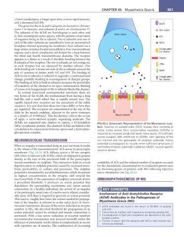
AChRs are organized into clusters by rapsyn, a 43-kilodalton FIG 65.3 Schematic Representation of the Myoneural Junc-
(kDa) cytoplasmic protein. The clustered AChRs are linked to the tion. Vesicles of acetylcholine (ACh) release their contents at
cytoskeleton by connections between rapsyn and a dystrophin– active zones across from acetylcholine receptors (AChRs) in
glycoprotein complex. response to impulses conducted down nerve axons. ACh diffuses
across synaptic cleft and binds to AChRs, with opening of the
NEUROMUSCULAR TRANSMISSION ion channel and the generation of endplate potential. Action
potential is propagated to muscle when sufficient amplitude of
When an impulse is transmitted along an axon terminal, it results summated endplate potentials is attained. MuSK, muscle-specific
in the release of the neurotransmitter ACh across its presynaptic tyrosine kinase.
membrane (Fig. 65.3). ACh diffuses across a 50-nm synaptic
cleft, where it interacts with AChRs, which are displayed in greatest
density at the tops of the junctional folds of the postsynaptic
muscle membrane or endplate. This interaction leads to a local availability of ACh and the reduced number of receptors accounts
depolarization or endplate potential caused by increased mem- for the characteristic decremental nerve conduction pattern seen
brane permeability to sodium and potassium. The endplate on electromyograms of patients with MG following repetitive
potential is terminated by acetylcholinesterases, which are present nerve stimulation (see Fig. 65.1).
in highest concentrations in the synaptic cleft around the
junctional folds. If the summation of endplate potentials attains IMMUNOPATHOGENESIS OF MG
a prescribed threshold, it produces an action potential that
depolarizes the surrounding sarcolemma and causes muscle
contraction. In a healthy individual, the arrival of an impulse KEY CONCEPtS
at the presynaptic membrane of a motor nerve releases consider- Involvement of Anti–Acetylcholine Receptor
ably more ACh than is required to generate an action potential. (AChR) Antibodies in the Pathogenesis of
This reserve, roughly four times the current needed for propaga-
tion of the impulse, is referred to as the safety factor of neuro- Myasthenia Gravis (MG)
muscular transmission. Because of the severe reduction in receptor • AChR antibodies are found in the serum of 85–90% of patients
number in MG, the electrical threshold for propagation of an with MG.
action potential cannot be attained and muscle contraction is • Infants born to mothers with myasthenia sometimes develop MG.
prevented. With a less severe reduction in receptor numbers • Immunoglobulin G (IgG) and complement are deposited at the post-
neuromuscular transmission may proceed normally unless the synaptic junction.
efficiency of presynaptic vesicle release is compromised, as occurs • Transfer of serum IgG from patients with MG to mice induces neu-
romuscular blockade.
with repetitive use of muscles. The combination of decreasing