
Page 1134 - Hall et al (2015) Principles of Critical Care-McGraw-Hill
P. 1134
CHAPTER 84: Cerebrovascular Disease 773
of patients with cerebrovascular disease remains to be demonstrated. Alternating pressure antithrombotic stockings may provide benefit as
Since the diagnosis of cerebral infarction can be made reliably by means well. In the case of pulmonary embolism or deep venous thrombosis, full
of the clinical picture and a CT scan, it is rarely if ever necessary to anticoagulation with heparin or heparin-like drugs may be instituted.
demonstrate a defect on a CBF study. Furthermore, other conditions Fever may occur due to infection or other systemic causes. Central
also may produce focal regional reductions of CBF. CBF measurement fevers due to hypothalamic disease are an exceedingly uncommon event
as an adjunct in deciding the appropriate therapeutic intervention in and the search for other causes should be vigorously pursued. Animal
patients with stroke has not been shown to result in improved outcome. studies have shown that even minor elevations in temperature of a few
The combination of diffusion weighted imaging (DWI) and perfusion degrees poststroke can lead to worse brain damage. Maintaining nor-
weighted imaging (PWI) in patients with acute ischemic stroke often mothermia through the use of antipyretics and cooling blankets makes
reveals a central area of restricted diffusion surrounded by a larger area good sense but is of unproven value. Trials of induced hypothermia with
of low perfusion. The diffusion abnormality increases with time and its both external and internal cooling are now underway. It is important
final boundaries correspond closely to the eventual infarct. These obser- to remember that dysphagia occurs commonly, even with unilateral
vations have led to the hypothesis that the area of perfusion-diffusion hemispheric lesions. Before oral feeding is instituted, each patient’s
mismatch indicates tissue destined for infarction that may be salvaged ability to swallow should be carefully checked. Institutions with formal
by thrombolytic therapy. As of 2012, several clinical trials all have failed dysphagia screening protocols have a reduced incidence of pneumonia.
31
to demonstrate that treatment that decisions based on DWI-PWI mag- Incontinence is also common following acute stroke but the use of Foley
netic resonance scans lead to better patient outcome. 22 catheters should be kept to a minimum because of the attendant increase
in urinary tract infections. Careful attention must be given to the pre-
vention of decubitus ulcers in bedridden patients.
TREATMENT Intravenously administered t-PA improves outcome in carefully
■ CEREBRAL INFARCTION selected patients with acute ischemic stroke when instituted within
4.5 hours of onset. These findings were demonstrated in two separate
5,6
Immediate supportive care of the patient with cerebral infarction requires studies: the NINDS Trial comprising patients within 0 to 3 hours of
attention to the patient’s airway, breathing, and circulation. Although onset and the ECASS III Trial comprising patients within 3 to 4.5 hours
most patients have preserved pharyngeal reflexes, those with brain stem of onset. Inclusion and exclusion criteria used in these trials were dif-
infarction or depressed consciousness may require intubation for airway ferent and are listed in Tables 84-1 and 84-2. In both trials, patients
protection. Coexisting heart and lung disease are common. Respiratory
and cardiac function should be assessed fully, and appropriate interven-
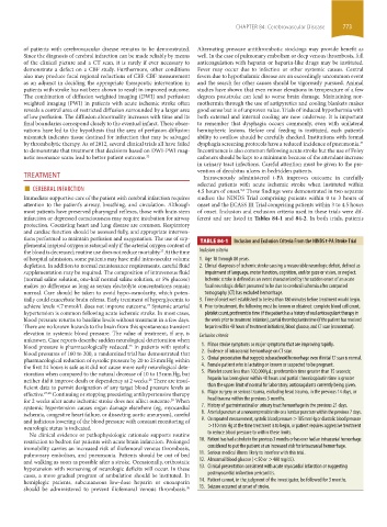
tions performed to maintain perfusion and oxygenation. The use of sup- TABLE 84-1 Inclusion and Exclusion Criteria From the NINDS t-PA Stroke Trial
plemental inspired oxygen is rational only if the arterial oxygen content of
the blood is decreased; routine use does not reduce mortality. At the time Inclusion criteria
23
of hospital admission, some patients may have mild intravascular volume 1. Age 18 through 80 years.
depletion. In addition to normal maintenance requirements, careful fluid 2. Clinical diagnosis of ischemic stroke causing a measurable neurologic deficit, defined as
supplementation may be required. The composition of intravenous fluid impairment of language, motor function, cognition, and/or gaze or vision, or neglect.
(normal saline solution, one-half normal saline solution, or 5% glucose) Ischemic stroke is defined as an event characterized by the sudden onset of an acute
makes no difference as long as serum electrolyte concentrations remain focal neurologic deficit presumed to be due to cerebral ischemia after computed
normal. Care should be taken to avoid hypo-osmolarity, which poten- tomography (CT) has excluded hemorrhage.
tially could exacerbate brain edema. Early treatment of hyperglycemia to 3. Time of onset well established to be less than 180 minutes before treatment would begin.
achieve levels <7 mmol/L does not improve outcome. Systemic arterial 4. Prior to treatment, the following must be known or obtained: complete blood cell count,
24
hypertension is common following acute ischemic stroke. In most cases, platelet count, prothrombin time (if the patient has a history of oral anticoagulant therapy in
blood pressure returns to baseline levels without treatment in a few days. the week prior to treatment initiation), partial thromboplastin time (if the patient has received
There are no known hazards to the brain from this spontaneous transient heparin within 48 hours of treatment initiation), blood glucose, and CT scan (noncontrast).
elevation in systemic blood pressure. The value of treatment, if any, is Exclusion criteria
unknown. Case reports describe sudden neurological deterioration when
blood pressure is pharmacologically reduced. In patients with systolic 1. Minor stroke symptoms or major symptoms that are improving rapidly.
25
blood pressures of 160 to 200, a randomized trial has demonstrated that 2. Evidence of intracranial hemorrhage on CT scan.
pharmacological reduction of systolic pressure by 20 to 25 mm Hg within 3. Clinical presentation that suggests subarachnoid hemorrhage even if initial CT scan is normal.
the first 24 hours is safe as it did not cause more early neurological dete- 4. Female patient who is lactating or known or suspected to be pregnant.
rioration when compared to the natural decrease of 10 to 15 mm Hg, but 5. Platelet count less than 100,000/µL; prothrombin time greater than 15 seconds;
neither did it improve death or dependency at 2 weeks. There are insuf- heparin has been given within 48 hours and partial thromboplastin time is greater
26
ficient data to permit designation of any target blood pressure levels as than the upper limit of normal for laboratory; anticoagulants currently being given.
effective. 27,28 Continuing or stopping preexisting antihypertensive therapy 6. Major surgery or serious trauma, excluding head trauma, in the previous 14 days, or
for 2 weeks after acute ischemic stroke does not affect outcome. When head trauma within the previous 3 months.
29
systemic hypertension causes organ damage elsewhere (eg, myocardial 7. History of gastrointestinal or urinary tract hemorrhage in the previous 21 days.
ischemia, congestive heart failure, or dissecting aortic aneurysm), careful 8. Arterial puncture at a noncompressible site or a lumbar puncture within the previous 7 days.
and judicious lowering of the blood pressure with constant monitoring of 9. On repeated measurement, systolic blood pressure >185 mm Hg or diastolic blood pressure
neurologic status is indicated. >110 mm Hg at the time treatment is to begin, or patient requires aggressive treatment
No clinical evidence or pathophysiologic rationale supports routine to reduce blood pressure to within these limits.
restriction to bedrest for patients with acute brain infarction. Prolonged 10. Patient has had a stroke in the previous 3 months or has ever had an intracranial hemorrhage
immobility carries an increased risk of iliofemoral venous thrombosis, considered to put the patient at an increased risk for intracranial hemorrhage.
pulmonary embolism, and pneumonia. Patients should be out of bed 11. Serious medical illness likely to interfere with this trial.
and walking as soon as possible after a stroke. Occasionally, orthostatic 12. Abnormal blood glucose (<50 or >400 mg/dL).
hypotension with worsening of neurologic deficits will occur. In these 13. Clinical presentation consistent with acute myocardial infarction or suggesting
cases, a more gradual program of ambulation should be instituted. In postmyocardial infarction pericarditis.
hemiplegic patients, subcutaneous low-dose heparin or enoxaparin 14. Patient cannot, in the judgment of the investigator, be followed for 3 months.
should be administered to prevent iliofemoral venous thrombosis. 15. Seizure occurred at onset of stroke.
30
section06.indd 773 1/23/2015 12:55:32 PM