
Page 1129 - Hall et al (2015) Principles of Critical Care-McGraw-Hill
P. 1129
768 PART 6: Neurologic Disorders
reduced or absent. In patients with CIP, direct needle stimulation of are short in duration, low in amplitude, and may be polyphasic. In
40
muscle elicits a relatively higher amplitude response compared with the contrast, long-duration, polyphasic, high amplitude MUPs may suggest
response recorded from muscle after nerve stimulation. neuropathy.
The serum CK level is normal, and, when performed, cerebrospinal Direct muscle stimulation can be conducted to assess for electrical
fluid protein levels are usually normal. Muscle biopsy findings are those inexcitability and may help to differentiate CIM from motor axonopa-
of neurogenic atrophy. Nerve histology in patients with electrophysi- thy. However, this modality is often limited to those patients with a
26
ologic-defined CIP demonstrates distal axonal degeneration involving coexisting peripheral neuropathy.
both sensory and motor fibers with no evidence of demyelination or Alternatively, CIM may be established with muscle biopsy. The major
inflammation. histopathologic finding is the selective loss of myosin, identified as a
Prior investigations have commonly associated CIP with severe lack of reactivity to myosin ATPase in non-necrotic fibers. This finding
sepsis and experts suspect it represents a neurologic manifestation of can be confirmed with immunohistochemic studies for myosin and by
the systemic inflammatory response syndrome (SIRS). 28,32,33 There is utilizing electron microscopy to identify loss of thick filaments. There
some correlation with elevations in blood glucose and reductions in is usually atrophy of myofibers (type 2 more than type 1), evidence of
serum albumin. The mechanism of axonal injury in CIP is unknown. myofibrillar disorganization, and occasional necrosis. 41,42
34
However, injury to the microcirculation of distal nerves, causing isch- Several processes may be involved in the pathogenesis of CIM, includ-
emia and axonal degeneration, is speculated. 33,35 During the early stages ing upregulation of calpain, an increase in muscle apoptosis, activation
of sepsis, electrical inexcitability due to sodium channel inactivation of the proteasome ubiquitin-degradative system, and upregulation of
may be present in otherwise intact nerves. the transforming growth factor-beta/mitogen-activated protein kinase
■ CRITICAL ILLNESS MYOPATHY (SEE TABlE 83-5) pathway. Oxidative stress may also play a role. Observation of the loss
43
of sarcolemmal nitric oxide synthase isoform 1 may lead to muscle fiber
The most common form of intensive care unit (ICU)-acquired myopa- inexcitability. 44
thy is critical illness myopathy (CIM). The most common presenting A steroid-denervation animal model reproduces the histopathologic
36
45
features of CIM are flaccid quadriparesis that may have a different pat- and electrophysiologic findings of CIM observed in humans. This
tern than CIP. Whereas CIP exhibits a length-related pattern (ie, distal model suggests that a deleterious interaction between glucocorticoids
muscles are weakest), CIM usually affects proximal muscles either and denervation leads to depletion of the mRNA for myosin and results
46
equally or more pronounced than distal muscles. Facial muscle weak- in muscle atrophy. Finally, muscle sodium channel properties have
ness can occur, but extraocular muscle weakness is rare. Like other also been implicated using a chronic sepsis animal model. Patch clamp
entities, patients often repeatedly fail to wean from mechanical ventila- technique revealed decreased sodium current that could lead to muscle
tion. Although not always assessable, sensation should be normal. For inexcitability. 47
example, these patients often grimace to painful stimuli even during ■
periods of delirium. CRITICAL ILLNESS POLYNEUROMYOPATHY
In retrospective series of patients with CIM, approximately one-half More recent investigations have proven that a reasonable proportion of
had elevations in CK. In patients with appropriate clinical features, the patients have features of combined CIM and CIP, termed critical illness
37
48
diagnosis of CIM can be confirmed by electrophysiologic testing with polyneuromyopathy. The commonality of this entity was illustrated by
nerve conduction studies (NCS) and electromyography (EMG). Muscle a prospective longitudinal cohort study of 48 patients who had baseline
biopsy establishes the diagnosis, but is rarely performed unless another neurologic examinations and nerve conduction studies (NCS) within
treatable condition, such as an inflammatory myopathy, is in the dif- 72 hours of developing severe sepsis. Electromyography was performed
49
ferential diagnosis. on patients who developed clinical weakness or had 30% or greater
The major nerve conduction findings of CIM are normal to low reduction in nerve conduction response amplitudes. Clinical and elec-
motor amplitudes with occasional broadening of the CMAP. 38,39 Phrenic trophysiologic examinations were repeated weekly for the duration of
motor amplitudes may also be low. Sensory responses are normal or the ICU stay. Abnormal NCS were present at baseline in 63% of patients,
only mildly reduced, unless there is a coexisting polyneuropathy. Needle and an abnormality on baseline NCS was significantly associated with
examination frequently demonstrates fibrillation potential activity hospital mortality compared with a normal baseline NCS (55% vs 0%,
implicating recent denervation or muscle necrosis. Observation of the respectively). In 20 patients who remained in the ICU long enough to
31
recruitment of motor unit potentials (MUPs) may not be possible in have serial NCS, neuromuscular dysfunction developed in 10 patients
advanced weakness. When feasible, recruitment tends to be early. MUPs (50%). Electrophysiologic evidence of both CIM and CIP was present
in 8 of 10 patients with neuromuscular dysfunction. The investigators
hypothesized that sepsis may be a common pathologic mechanism
underlying the development of both CIM and CIP.
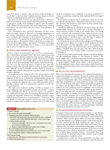
TABLE 83-5 Major Diagnostic Features of CIM ■ PROLONGED NEUROMUSCULAR JUNCTION BLOCKADE
1. Evidence for ICUAW
2. Intact sensory examination (when possible) Prolonged neuromuscular junction (NMJ) blockade is a rare disorder
3. Electrophysiologic evidence of myopathy without neuropathy occurring in patients who receive non-depolarizing NMBAs who experi-
a. Needle EMG with short-duration, low-amplitude MUPs with early or normal full ence persistent generalized weakness and respiratory failure despite drug
50
recruitment, with or without fibrillation potentials in 2 or more muscle groups cessation. These paralytic agents inhibit neuromuscular transmission
b. Absence of other nerve injury via reversible binding to acetylcholine receptors on the motor end-plates
i. Sensory nerve amplitudes >80% of the lower limit of normal in two or more of NMJs. However, specific drugs requiring end organ function for
nerves on nerve conduction studies clearance may have persistent effects, particularly when infused for pro-
ii. Absence of a decremental response on repetitive nerve stimulation longed periods. For example, aminosteroid blocking agents, such as pan-
4. Muscle inexcitability on direct muscle stimulation curonium and vecuronium, undergo metabolism by the liver and result
a. Muscle-stimulated CMAP/nerve-evoked CMAP ratio >0.5 in 2 or more muscles in functionally active 3-hydroxy metabolites. In situations of advanced
5. Muscle histopathologic findings of myopathy with myosin loss liver or kidney injury ( creatinine clearance <30 mL/min), these drugs
can accumulate for prolonged effect. Other reported contexts include
Other supportive findings: hypermagnesemia or metabolic acidosis.
1. Motor amplitudes <80% of the lower limit of normal in two or more nerves Examination is notable for flaccid quadriplegia, arreflexia, and
2. Elevated serum creatine kinase involvement of the cranial nerves, including ptosis, ophthalmoparesis,
section06.indd 768 1/23/2015 12:55:31 PM