
Page 135 - Review of Medical Microbiology and Immunology ( PDFDrive )
P. 135
mebooksfree.com
mebooksfree.com
mebooksfree.com
mebooksfree.com
mebooksfree.com
mebooksfree.com
mebooksfree.com
mebooksfree.com mebooksfree.com mebooksfree.com to be a better predictor of heart attack risk than an elevated mebooksfree.com
mebooksfree.com
PART II Clinical Bacteriology
124
cholesterol level.
Transmission
Humans are the natural hosts for pneumococci; there is no
animal reservoir. Because a proportion (5%–50%) of the
healthy population harbors virulent organisms in the oro-
pharynx, pneumococcal infections are not considered to be
communicable. Resistance is high in healthy young people,
mebooksfree.com mebooksfree.com mebooksfree.com Pathogenesis mebooksfree.com mebooksfree.com
mebooksfree.com
and disease results most often when predisposing factors
(see following discussion) are present.
The most important virulence factor is the capsular poly-
saccharide, and anticapsular antibody is protective. Lipo-
teichoic acid, which activates complement and induces
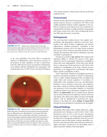
FIGURE 15–15
Streptococcus pneumoniae—Gram stain.
inflammatory cytokine production, contributes to the
Arrows point to typical gram-positive diplococci. Note that the clear
inflammatory response and to the septic shock syndrome
area around the organism is the capsule. (Used with permission from Pro-
that occurs in some immunocompromised patients. Pneu-
fessor Shirley Lowe, University of California, San Francisco School of Medicine.)
molysin, the hemolysin that causes α-hemolysis, may also
contribute to pathogenesis.
Pneumococci produce IgA protease that enhances the
mebooksfree.com
mebooksfree.com mebooksfree.com mebooksfree.com there is outpouring of fluid and red and white blood cells, mebooksfree.com
mebooksfree.com
α-, β-, and γ-globulins.) Note that CRP is a nonspecific
organism’s ability to colonize the mucosa of the upper
indicator of inflammation and is elevated in response to
respiratory tract by cleaving IgA. Pneumococci multiply in
the presence of many organisms, not just S. pneumoniae.
tissues and cause inflammation. When they reach alveoli,
Clinically, CRP in human serum is measured in the laboratory
by its reaction with the carbohydrate of S. pneumoniae. The
resulting in consolidation of the lung. During recovery,
medical importance of CRP is that an elevated CRP appears
pneumococci are phagocytized, mononuclear cells ingest
debris, and the consolidation resolves.
Factors that lower resistance and predispose persons to
pneumococcal infection include (1) alcohol or drug intoxi-
cation or other cerebral impairment that can depress the
cough reflex and increase aspiration of secretions; (2)
abnormality of the respiratory tract (e.g., viral infections),
pooling of mucus, bronchial obstruction, and respiratory
mebooksfree.com
mebooksfree.com
mebooksfree.com mebooksfree.com mebooksfree.com failure); (4) splenectomy; and (5) certain chronic diseases mebooksfree.com
tract injury caused by irritants (which disturb the integrity
and movement of the mucociliary blanket); (3) abnormal
circulatory dynamics (e.g., pulmonary congestion and heart
such as sickle cell anemia and nephrosis. Patients with sickle
cell anemia auto-infarct their spleen, become functionally
asplenic, and are predisposed to pneumococcal sepsis.
Trauma to the head that causes leakage of spinal fluid
through the nose predisposes to pneumococcal meningitis.
FIGURE 15–16
Optochin test—Arrow points to zone of inhi-
Pneumonia often begins with a sudden chill, fever, cough,
bition of growth of Streptococcus pneumoniae caused by optochin Clinical Findings
and pleuritic pain. Sputum is a red or brown “rusty” color.
that has diffused from the disk labeled P. In the lower half of the
mebooksfree.com
mebooksfree.com mebooksfree.com mebooksfree.com prominent cause of otitis media, sinusitis, mastoiditis, con- mebooksfree.com
mebooksfree.com
Bacteremia occurs in 15% to 25% of cases. Spontaneous
blood agar plate, there is α-hemolysis caused by S. pneumoniae,
recovery may begin in 5 to 10 days and is accompanied by
except in the region around the optochin disk. The arrow points to
development of anticapsular antibodies. Pneumococci are a
the outer limit of the zone of inhibition. Upper half of blood agar
plate shows α-hemolysis caused by a viridans streptococcus, and
junctivitis, purulent bronchitis, pericarditis, bacterial men-
there is no zone of inhibition around the optochin disk. (Used with per-
ingitis, and sepsis. Pneumococci are the leading cause of
mission from Professor Shirley Lowe, University of California, San Francisco School of
sepsis in patients without a functional spleen.
Medicine.)
mebooksfree.com mebooksfree.com mebooksfree.com mebooksfree.com mebooksfree.com mebooksfree.com