
Page 212 - Review of Medical Microbiology and Immunology ( PDFDrive )
P. 212
mebooksfree.com
mebooksfree.com
mebooksfree.com
mebooksfree.com
mebooksfree.com
mebooksfree.com mebooksfree.com mebooksfree.com mebooksfree.com mebooksfree.com mebooksfree.com
mebooksfree.com
201
CHAPTER 24 Spirochetes
mebooksfree.com mebooksfree.com mebooksfree.com mebooksfree.com mebooksfree.com mebooksfree.com
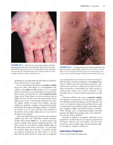
mebooksfree.com mebooksfree.com mebooksfree.com FIGURE 24–4 Condylomata lata of secondary syphilis. Note the mebooksfree.com
mebooksfree.com
mebooksfree.com
FIGURE 24–3
Palmar lesions of secondary syphilis. Note the
papulosquamous lesions on the right palm. Palmar lesions are typi-
cally bilateral. (Reproduced with permission from Wolff K, Johnson R. Fitzpatrick’s
flat, moist perianal lesions (black arrow). (Reproduced with permission from
Color Atlas & Synopsis of Clinical Dermatology. 6th ed. New York: McGraw-Hill, 2009.
Wolff K, Johnson R. Fitzpatrick’s Color Atlas & Synopsis of Clinical Dermatology. 6th ed.
Copyright © 2009 by The McGraw-Hill Companies, Inc.)
New York: McGraw-Hill, 2009. Copyright © 2009 by The McGraw-Hill Companies, Inc.)
occur and patients are not infectious. In the remaining one-
spontaneously, but spirochetes spread widely via the blood-
third of people, the disease progresses to the tertiary stage.
stream (bacteremia) to many organs.
One to 3 months later, the lesions of secondary syphilis
cially of skin and bones; central nervous system involve-
may occur. These often appear as a maculopapular rash,
ment, also known as neurosyphilis (e.g., tabes, paresis); or
notably on the palms and soles (Figure 24–3), or as moist Tertiary syphilis may show granulomas (gummas), espe-
cardiovascular lesions (e.g., aortitis, aneurysm of the
mebooksfree.com mebooksfree.com mebooksfree.com The organism is transmitted across the placenta, typically mebooksfree.com
mebooksfree.com
mebooksfree.com
papules on skin and mucous membranes (mucous patches).
ascending aorta). In tertiary lesions, treponemes are rarely
Moist lesions on the genitals are called condylomata lata
seen.
(Figure 24–4). These lesions are rich in spirochetes and are
Treponema pallidum also causes congenital syphilis.
highly infectious, but they also heal spontaneously. Patchy
alopecia also occurs. Constitutional symptoms of second-
after the third month of pregnancy, and fetal infection can
ary syphilis include low-grade fever, malaise, anorexia,
occur. In the infected neonates, skin and bone lesions, such
weight loss, headache, myalgias, and generalized lymph-
as Hutchinson’s teeth, mulberry molars, saber shins, saddle
adenopathy. Pharyngitis, meningitis, nephritis, and hepati-
nose, rhagades, snuffles, and frontal bossing, are common.
tis may also occur. In some individuals, the symptoms of
Other findings, such as hepatosplenomegaly, interstitial
the primary and secondary stages may not occur, and yet
the disease may progress.
tion can also result in stillbirth.
About one-third of these early (primary and secondary)
Immunity to syphilis is incomplete. Antibodies to the
syphilis cases will “cure” themselves, without treatment. keratitis, and eighth nerve deafness, also occur. Fetal infec-
organism are produced but do not stop the progression of
mebooksfree.com
mebooksfree.com mebooksfree.com mebooksfree.com lis are relatively resistant to reinfection. mebooksfree.com mebooksfree.com
Another third remain latent (i.e., no lesions appear, but
the disease. Patients with early syphilis who have been
positive serologic tests indicate continuing infection). The
treated can contract syphilis again. Patients with late syphi-
latent period can be divided into early and late stages. In
the early latent period, which can last for 1 or 2 years after
the secondary stage, the symptoms of secondary syphilis
Laboratory Diagnosis
can reappear and patients can infect others. In the late
latent period, which can last for many years, no symptoms
There are three important approaches.
mebooksfree.com mebooksfree.com mebooksfree.com mebooksfree.com mebooksfree.com mebooksfree.com