
Page 317 - Review of Medical Microbiology and Immunology ( PDFDrive )
P. 317
mebooksfree.com
mebooksfree.com
mebooksfree.com
mebooksfree.com
mebooksfree.com
mebooksfree.com
mebooksfree.com
mebooksfree.com mebooksfree.com mebooksfree.com produced by squamous cells on the surface, which enhances mebooksfree.com
mebooksfree.com
PART IV Clinical Virology
306
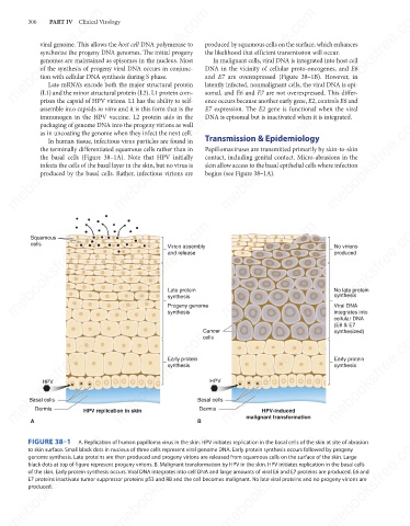
viral genome. This allows the host cell DNA polymerase to
synthesize the progeny DNA genomes. The initial progeny
the likelihood that efficient transmission will occur.
In malignant cells, viral DNA is integrated into host cell
genomes are maintained as episomes in the nucleus. Most
DNA in the vicinity of cellular proto-oncogenes, and E6
of the synthesis of progeny viral DNA occurs in conjunc-
tion with cellular DNA synthesis during S phase.
and E7 are overexpressed (Figure 38–1B). However, in
Late mRNA’s encode both the major structural protein
somal, and E6 and E7 are not overexpressed. This differ-
(L1) and the minor structural protein (L2). L1 protein com-
ence occurs because another early gene, E2, controls E6 and
prises the capsid of HPV virions. L1 has the ability to self-
E7 expression. The E2 gene is functional when the viral
assemble into capsids in vitro and it is this form that is the latently infected, nonmalignant cells, the viral DNA is epi-
immunogen in the HPV vaccine. L2 protein aids in the
mebooksfree.com mebooksfree.com mebooksfree.com Transmission & Epidemiology mebooksfree.com mebooksfree.com
DNA is episomal but is inactivated when it is integrated.
mebooksfree.com
packaging of genome DNA into the progeny virions as well
as in uncoating the genome when they infect the next cell.
In human tissue, infectious virus particles are found in
Papillomaviruses are transmitted primarily by skin-to-skin
the terminally differentiated squamous cells rather than in
the basal cells (Figure 38–1A). Note that HPV initially
contact, including genital contact. Micro-abrasions in the
skin allow access to the basal epithelial cells where infection
infects the cells of the basal layer in the skin, but no virus is
produced by the basal cells. Rather, infectious virions are
begins (see Figure 38–1A).
mebooksfree.com mebooksfree.com mebooksfree.com mebooksfree.com mebooksfree.com mebooksfree.com
Squamous
cells
No virions
Virion assembly
and release
produced
Late protein
No late protein
synthesis
synthesis
Viral DNA
Progeny genome
synthesis
integrates into
cellular DNA
(E6 & E7
mebooksfree.com mebooksfree.com mebooksfree.com HPV mebooksfree.com mebooksfree.com mebooksfree.com
Cancer
synthesized)
cells
Early protein
Early protein
synthesis
synthesis
HPV
Basal cells
Basal cells
Dermis
Dermis
HPV replication in skin
HPV-induced
malignant transformation
B
A
mebooksfree.com mebooksfree.com mebooksfree.com mebooksfree.com mebooksfree.com mebooksfree.com
FIGURE 38–1
A. Replication of human papilloma virus in the skin. HPV initiates replication in the basal cells of the skin at site of abrasion
to skin surface. Small black dots in nucleus of three cells represent viral genome DNA. Early protein synthesis occurs followed by progeny
genome synthesis. Late proteins are then produced and progeny virions are released from squamous cells on the surface of the skin. Large
black dots at top of figure represent progeny virions. B. Malignant transformation by HPV in the skin. HPV initiates replication in the basal cells
of the skin. Early protein synthesis occurs. Viral DNA integrates into cell DNA and large amounts of viral E6 and E7 proteins are produced. E6 and
E7 proteins inactivate tumor suppressor proteins p53 and RB and the cell becomes malignant. No late viral proteins and no progeny virions are
produced.
mebooksfree.com mebooksfree.com mebooksfree.com mebooksfree.com mebooksfree.com mebooksfree.com