
Page 416 - Review of Medical Microbiology and Immunology ( PDFDrive )
P. 416
mebooksfree.com
mebooksfree.com
mebooksfree.com
mebooksfree.com
mebooksfree.com
mebooksfree.com
mebooksfree.com
mebooksfree.com mebooksfree.com mebooksfree.com In human immunodeficiency virus (HIV)–infected 405 mebooksfree.com
mebooksfree.com
CHAPTER 48 Cutaneous & Subcutaneous Mycoses
Tinea Versicolor
patients with low CD4 counts, disseminated sporotrichosis
Tinea versicolor (pityriasis versicolor), a superficial skin
can occur. Sporotrichosis occurs most often in gardeners,
infection of cosmetic importance only, is caused by Malas-
especially those who prune roses, because they may be
sezia species. The lesions are usually noticed as hypopig-
stuck by a rose thorn.
mented areas, especially on tanned skin in the summer.
In the clinical laboratory, round or cigar-shaped bud-
There may be slight scaling or itching, but usually the infec-
tion is asymptomatic. It occurs more frequently in hot,
temperature, hyphae occur bearing oval conidia in clusters
humid weather. The lesions contain both budding yeast
at the tip of slender conidiophores (resembling a daisy).
cells and hyphae. Diagnosis is usually made by observing ding yeasts are seen in tissue specimens. In culture at room
mebooksfree.com
mebooksfree.com mebooksfree.com mebooksfree.com Chromomycosis mebooksfree.com mebooksfree.com
The drug of choice for skin lesions is itraconazole (Spo-
this mixture in KOH preparations of skin scrapings. Cul-
ranox). It can be prevented by protecting skin when touch-
ture is not usually done. The treatment of choice is topical
ing plants, moss, and wood.
miconazole, but the lesions have a tendency to recur. Oral
antifungal drugs, such as fluconazole or itraconazole, can
be used to treat recurrences.
This is a slowly progressive granulomatous infection that is
caused by several soil fungi (Fonsecaea, Phialophora, Clado-
Tinea Nigra
sporium, etc.) when introduced into the skin through
Tinea nigra is an infection of the keratinized layers of the
trauma. These fungi are collectively called dematiaceous
skin. It appears as a brownish spot caused by the melanin-
like pigment in the hyphae. The causative organism, Clado-
colored, either gray or black. Wartlike lesions with crusting
sporium werneckii, is found in the soil and transmitted
abscesses extend along the lymphatics. The disease occurs
during injury. In the United States, the disease is seen in the fungi, so named because their conidia or hyphae are dark-
mainly in the tropics and is found on bare feet and legs. In
southern states. Diagnosis is made by microscopic exami-
mebooksfree.com
mebooksfree.com mebooksfree.com mebooksfree.com oral flucytosine or thiabendazole, plus local surgery. mebooksfree.com
mebooksfree.com
the clinical laboratory, dark brown, round fungal cells are
nation and culture of skin scrapings. The infection is
seen in leukocytes or giant cells. The disease is treated with
treated with a topical keratolytic agent (e.g., salicylic acid).
SUBCUTANEOUS MYCOSES
Mycetoma
Soil fungi (Petriellidium, Madurella) enter through wounds
These are caused by fungi that grow in soil and on vegeta-
on the feet, hands, or back and cause abscesses, with pus
tion and are introduced into subcutaneous tissue through
discharged through sinuses. The pus contains compact
trauma.
colored granules. Actinomycetes such as Nocardia can
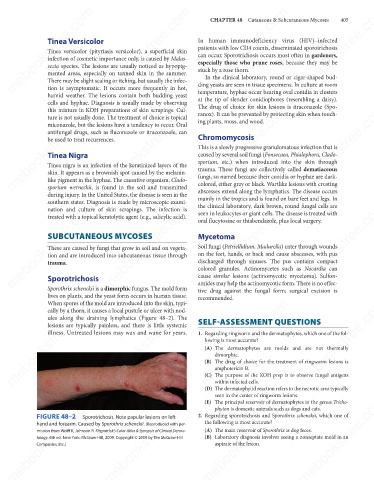
Sporotrichosis
amides may help the actinomycotic form. There is no effec-
Sporothrix schenckii is a dimorphic fungus. The mold form
tive drug against the fungal form; surgical excision is
lives on plants, and the yeast form occurs in human tissue. cause similar lesions (actinomycotic mycetoma). Sulfon-
recommended.
mebooksfree.com mebooksfree.com mebooksfree.com SELF-ASSESSMENT QUESTIONS mebooksfree.com
mebooksfree.com
mebooksfree.com
When spores of the mold are introduced into the skin, typi-
cally by a thorn, it causes a local pustule or ulcer with nod-
ules along the draining lymphatics (Figure 48–2). The
lesions are typically painless, and there is little systemic
illness. Untreated lesions may wax and wane for years.
1. Regarding ringworm and the dermatophytes, which one of the fol-
lowing is most accurate?
(A) The dermatophytes are molds and are not thermally
dimorphic.
(B) The drug of choice for the treatment of ringworm lesions is
amphotericin B.
(C) The purpose of the KOH prep is to observe fungal antigens
within infected cells.
(D) The dermatophytid reaction refers to the necrotic area typically
seen in the center of ringworm lesions.
mebooksfree.com mebooksfree.com mebooksfree.com (A) The main reservoir of Sporothrix is dog feces. mebooksfree.com
mebooksfree.com
mebooksfree.com
(E) The principal reservoir of dermatophytes in the genus Tricho-
phyton is domestic animals such as dogs and cats.
FIGURE 48–2
2. Regarding sporotrichosis and Sporothrix schenckii, which one of
Sporotrichosis. Note papular lesions on left
the following is most accurate?
hand and forearm. Caused by Sporothrix schenckii. (Reproduced with per-
mission from Wolff K, Johnson R. Fitzpatrick’s Color Atlas & Synopsis of Clinical Derma-
(B) Laboratory diagnosis involves seeing a nonseptate mold in an
tology. 6th ed. New York: McGraw-Hill, 2009. Copyright © 2009 by The McGraw-Hill
aspirate of the lesion.
Companies, Inc.)
mebooksfree.com mebooksfree.com mebooksfree.com mebooksfree.com mebooksfree.com mebooksfree.com