
Page 465 - Review of Medical Microbiology and Immunology ( PDFDrive )
P. 465
mebooksfree.com
mebooksfree.com
mebooksfree.com
mebooksfree.com
mebooksfree.com
mebooksfree.com
mebooksfree.com
mebooksfree.com
mebooksfree.com mebooksfree.com Insect mebooksfree.com Stage(s) in Humans Most Important Stage(s) mebooksfree.com
PART VI Parasitology
454
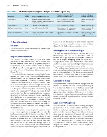
TABLE 54–2 Medically Important Stages in Life Cycle of Cestodes (Tapeworms)
Vector
Stage That Infects Humans
Associated with Disease
Outside of Humans
Organism
Taenia solium
Larvae in muscle of pig
None
1. Larvae in undercooked pork
Adult tapeworm in intestine
with human feces
Larvae in muscle of pig
Adult tapeworm in intestine
Larvae in undercooked beef
Taenia saginata
Diphyllobothrium latum None 2. Eggs in food or water contaminated Cysticercus, especially in brain None
Larvae in muscle of
None
Larvae in undercooked fish
Adult tapeworm in intestine can
cause vitamin B 12 deficiency
freshwater fish
mebooksfree.com
mebooksfree.com
mebooksfree.com mebooksfree.com mebooksfree.com vessel. They can disseminate to many organs, especially mebooksfree.com
Hydatid cysts, especially in liver and
None
Echinococcus granulosus
Eggs in food or water contaminated
Adult tapeworm in dog
lung
intestine produces
with dog feces
eggs
1. Taenia solium
the eyes and brain, where they encyst to form cysticerci
Disease
(Figure 54–6). Each cysticercus contains a larva.
The adult form of T. solium causes taeniasis. Taenia solium
larvae cause cysticercosis.
The adult tapeworm attached to the intestinal wall causes
little damage. The cysticerci, on the other hand, can
Important Properties Pathogenesis & Epidemiology
become very large, especially in the brain, where they
mebooksfree.com
mebooksfree.com
mebooksfree.com mebooksfree.com mebooksfree.com die, they can release substances that provoke an inflamma- mebooksfree.com
The life cycle of T. solium is shown in Figure 54–1. Taenia
manifest as a space-occupying lesion (see Figure 54–6).
solium can be identified by its scolex, which has four suck-
Living cysticerci do not cause inflammation, but when they
ers and circle of hooks, and by its gravid proglottids,
which have 5 to 10 primary uterine branches (Figures
tory response. Eventually, the cysticerci calcify.
54–2A, B and 54–3). The eggs appear the same micro-
The epidemiology of taeniasis and cysticercosis is related
scopically as those of T. saginata and Echinococcus species
to the access of pigs to human feces and to consumption of
(Figure 54–4A).
raw or undercooked pork. The disease occurs worldwide
In taeniasis, the adult tapeworm is located in the human
but is endemic in areas of Asia, South America, and Eastern
intestine (see Figure 54–1). This occurs when humans are
Europe. Most cases in the United States are imported.
infected by eating raw or undercooked pork containing the
larvae, called cysticerci. (A cysticercus consists of a pea-
sized fluid-filled bladder with an invaginated scolex.) In the
Most patients with adult tapeworms are asymptomatic, but
small intestine, the larvae attach to the gut wall and take Clinical Findings
anorexia and diarrhea can occur. Some may notice proglot-
mebooksfree.com
mebooksfree.com mebooksfree.com mebooksfree.com appear as uveitis or retinitis, or the larvae can be visualized mebooksfree.com
mebooksfree.com
about 3 months to grow into adult worms measuring up to
tids in the stools. Cysticercosis in the brain causes head-
5 m. The gravid terminal proglottids containing many eggs
ache, vomiting, and seizures. Cysticercosis in the eyes can
detach daily, are passed in the feces, and are accidentally
eaten by pigs. Note that pigs are infected by the worm eggs;
floating in the vitreous. Subcutaneous nodules containing
therefore, it is the larvae (cysticerci) that are found in the
cysticerci commonly occur. Cysts also are commonly found
pig. A six-hooked embryo (oncosphere) emerges from each
in skeletal muscle.
egg in the pig’s intestine. The embryos burrow into a blood
vessel and are carried to skeletal muscle. They develop into
Laboratory Diagnosis
cysticerci in the muscle, where they remain until eaten by a
human. Humans are the definitive hosts, and pigs are the
glottids with 5 to 10 primary uterine branches in the
intermediate hosts.
In cysticercosis, a more dangerous sequence occurs
stools. In contrast, T. saginata proglottids have 15 to 20
when a person ingests the worm eggs in food or water that Identification of T. solium consists of finding gravid pro-
primary uterine branches. Eggs are found in the stools less
mebooksfree.com
mebooksfree.com
mebooksfree.com mebooksfree.com mebooksfree.com (CT) scan. Serologic tests (e.g., enzyme-linked immuno- mebooksfree.com
often than are proglottids. Diagnosis of cysticercosis
has been contaminated with human feces (Figure 54–5).
depends on demonstrating the presence of the cyst in tis-
Note that in cysticercosis, humans are infected by eggs
excreted in human feces, not by ingesting undercooked
sue, usually by surgical removal or computed tomography
pork. Also, pigs do not have the adult worm in their intes-
sorbent assay [ELISA]) that detect antibodies to T. solium
tine, so they are not the source of the eggs that cause
human cysticercosis. The eggs hatch in the small intestine,
antigens are available, but they may be negative in
and the oncospheres burrow through the wall into a blood
neurocysticercosis.
mebooksfree.com mebooksfree.com mebooksfree.com mebooksfree.com mebooksfree.com mebooksfree.com