
Page 476 - Review of Medical Microbiology and Immunology ( PDFDrive )
P. 476
mebooksfree.com
mebooksfree.com
mebooksfree.com
mebooksfree.com
mebooksfree.com
mebooksfree.com mebooksfree.com mebooksfree.com mebooksfree.com mebooksfree.com mebooksfree.com
mebooksfree.com
CHAPTER 55 Trematodes
465
B
C
mebooksfree.com mebooksfree.com mebooksfree.com mebooksfree.com mebooksfree.com mebooksfree.com
A
D
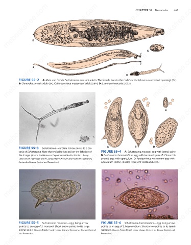
FIGURE 55–2
A: Male and female Schistosoma mansoni adults. The female lives in the male’s schist (shown as a ventral opening) (6×).
B: Clonorchis sinensis adult (6×). C: Paragonimus westermani adult (0.6×). D: S. mansoni cercaria (300×).
mebooksfree.com mebooksfree.com mebooksfree.com A mebooksfree.com Dmebooksfree.com mebooksfree.com
C
B
FIGURE 55–3
Schistosoma—cercaria. Arrow points to a cer-
caria of Schistosoma. Note the typical forked tail on the left side of FIGURE 55–4 A: Schistosoma mansoni egg with lateral spine.
mebooksfree.com mebooksfree.com mebooksfree.com mebooksfree.com mebooksfree.com mebooksfree.com
B: Schistosoma haematobium egg with terminal spine. C: Clonorchis
the image. (Source: the Minnesota Department of Health, R.N. Barr Library;
sinensis egg with operculum. D: Paragonimus westermani egg with
Librarians M. Rethlefson and M. Jones; Prof. W.Wiley, Public Health Image Library,
operculum (300×). (Circles represent red blood cells.)
Centers for Disease Control and Prevention.)
mebooksfree.com
mebooksfree.com
mebooksfree.com mebooksfree.com mebooksfree.com FIGURE 55–6 Schistosoma haematobium—egg. Long arrow mebooksfree.com
FIGURE 55–5
Schistosoma mansoni—egg. Long arrow
points to an egg of S. haematobium. Short arrow points to its termi-
points to an egg of S. mansoni. Short arrow points to its large
lateral spine. (Source: Public Health Image Library, Centers for Disease Control
nal spine. (Source: Public Health Image Library, Centers for Disease Control and
Prevention.)
and Prevention.)
mebooksfree.com mebooksfree.com mebooksfree.com mebooksfree.com mebooksfree.com mebooksfree.com