
Page 519 - Review of Medical Microbiology and Immunology ( PDFDrive )
P. 519
mebooksfree.com
mebooksfree.com
mebooksfree.com
mebooksfree.com
mebooksfree.com
mebooksfree.com
mebooksfree.com mebooksfree.com Antigen TCR Helper presenting Class II Antigen TCR Helper mebooksfree.com
mebooksfree.com
mebooksfree.com
mebooksfree.com
PART VII Immunology
508
Class II
MHC
Antigen-
Antigen-
MHC
presenting
cell
cell
CD28
B7 CD4 protein IL-2R T cell IL-2 protein CD4 protein T cell
B7
CTLA-4
protein
mebooksfree.com mebooksfree.com mebooksfree.com mebooksfree.com mebooksfree.com mebooksfree.com
protein
protein
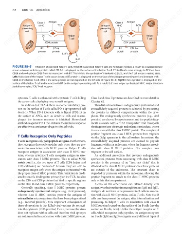
FIGURE 58–5
Inhibition of activated helper T cells. When the activated helper T cells are no longer needed, a return to a quiescent state
occurs when an inhibitory protein called CTLA-4 is displayed on the surface of the helper T cell. CTLA-4 binds more strongly to B7 than does
CD28 and so displaces CD28 from its interaction with B7. This inhibits the synthesis of interleukin-2 (IL-2), and the T cell enters a resting state.
Left: Activation of the helper T cells occurs because B7 protein is displayed on the surface of the antigen-presenting cell and interacts with
CD28 on the helper T cell. (This is the same process as that depicted on the left side of Figure 58–4.) Right: CTLA-4 protein is displayed on the
surface of the helper T cell and interacts with B7 on the antigen-presenting cell. As a result, IL-2 is no longer synthesized. MHC, major histocom-
patibility complex; TCR, T-cell receptor.
cytotoxic T cells is enhanced with cytotoxic T cells killing
mebooksfree.com
mebooksfree.com mebooksfree.com mebooksfree.com Class I and class II proteins are described in more detail in mebooksfree.com
mebooksfree.com
Chapter 62.
the cancer cells displaying new, nonself antigens.
In addition to CTLA-4, there is another inhibitory pro-
This distinction between endogenously synthesized and
extracellularly acquired proteins is achieved by processing
tein on the surface of T cells called PD-1 (programmed cell
the proteins in different compartments within the cyto-
death-1). When PD-1 interacts with its ligand (PDL-1) on
the surface of APCs, such as dendritic cells and macro-
plasm. The endogenously synthesized proteins (e.g., viral
phages, the immune response is inhibited. Monoclonal
proteins) are cleaved by a proteasome, and the peptide frag-
antibodies against PD-1 that enhance the immune response
ments associate with a “TAP transporter” that transports
are effective as anticancer drugs in clinical trials.
the fragment into the rough endoplasmic reticulum, where
it associates with the class I MHC protein. The complex of
T Cells Recognize Only Peptides
via the Golgi apparatus to the cell surface. In contrast, the
extracellularly acquired proteins are cleaved to peptide
T cells recognize only polypeptide antigens. Furthermore,
they recognize those polypeptides only when they are pre- peptide fragment and class I MHC protein then migrates
fragments within an endosome, where the fragment associ-
mebooksfree.com
mebooksfree.com mebooksfree.com mebooksfree.com synthesized proteins from associating with class II MHC mebooksfree.com
mebooksfree.com
ates with class II MHC proteins. This complex then
sented in association with MHC proteins. Helper T cells
recognize antigen in association with class II MHC pro-
migrates to the cell surface.
An additional protection that prevents endogenously
teins, whereas cytotoxic T cells recognize antigen in asso-
ciation with class I MHC proteins. This is called MHC
proteins is the presence of an “invariant chain” that is
restriction (i.e., the two types of T cells [CD4 helper and
attached to the class II MHC proteins when these proteins
CD8 cytotoxic] are “restricted” because they are able to
recognize antigen only when the antigen is presented with
are outside of the endosome. The invariant chain is
the proper class of MHC protein). This restriction is medi-
degraded by proteases within the endosome, allowing the
ated by specific binding sites primarily on the TCR, but also
peptide fragment to attach to the class II MHC proteins
on the CD4 and CD8 proteins that bind to specific regions
on the class II and class I MHC proteins, respectively.
B cells, on the other hand, can interact directly with
Generally speaking, class I MHC proteins present
antigens via their surface immunoglobulins (IgM and IgD).
Antigens do not have to be presented to B cells in associa-
endogenously synthesized antigens (e.g., viral proteins), only within that compartment.
mebooksfree.com
mebooksfree.com mebooksfree.com mebooksfree.com MHC proteins located on the surface of the B cells (see the mebooksfree.com
mebooksfree.com
whereas class II MHC proteins present the antigens of
tion with class II MHC proteins, unlike T cells. Note that B
cells can then present the antigen, after internalization and
extracellular microorganisms that have been phagocytized
processing, to helper T cells in association with class II
(e.g., bacterial proteins). One important consequence of
these observations is that killed viral vaccines do not acti-
vate the cytotoxic (CD8-positive) T cells, because the virus
section on B cells, later). Unlike the antigen receptor on T
does not replicate within cells and therefore viral epitopes
cells, which recognizes only peptides, the antigen receptors
on B cells (IgM and IgD) recognize many different types of
are not presented in association with class I MHC proteins.
mebooksfree.com mebooksfree.com mebooksfree.com mebooksfree.com mebooksfree.com mebooksfree.com