
Page 575 - Review of Medical Microbiology and Immunology ( PDFDrive )
P. 575
mebooksfree.com
mebooksfree.com
mebooksfree.com
mebooksfree.com
mebooksfree.com mebooksfree.com Macrophage IL-1, IL-12 Helper Tuberculin-Type Hypersensitivity mebooksfree.com mebooksfree.com
mebooksfree.com
mebooksfree.com
mebooksfree.com
mebooksfree.com
PART VII Immunology
564
Antigen
Delayed hypersensitivity to antigens of microorganisms
+
occurs in many infectious diseases and has been used as an
aid in diagnosis. It is typified by the tuberculin reaction.
Macrophage
When a patient previously exposed to Mycobacterium
T cell
(Th-1)
(purified protein derivative [PPD]) intradermally, there is
TCR
little reaction in the first few hours. Gradually, however,
Class II
MHC protein Gamma-interferon tuberculosis is injected with a small amount of tuberculin
induration and redness develop and reach a peak in 48 to
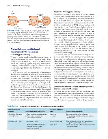
FIGURE 65–4
mebooksfree.com mebooksfree.com mebooksfree.com verts from negative to positive, it suggests that the patient mebooksfree.com
mebooksfree.com
mebooksfree.com
Delayed (cell-mediated) hypersensitivity. The
72 hours. A positive skin test indicates that the person has
macrophage ingests the antigen, processes it, and presents an
been infected with the agent, but it does not confirm the
epitope on its surface in association with class II major histocom-
presence of current disease. However, if the skin test con-
patibility complex (MHC) protein. The helper T (Th-1) cell is
activated and produces gamma interferon, which activates macro-
has been recently infected. Infected persons do not always
phages. These two types of cells mediate delayed hypersensitivity.
have a positive skin test, because overwhelming infection,
TCR, T-cell receptor.
disorders that suppress cell-mediated immunity (e.g., uremia,
measles, sarcoidosis, lymphoma, and acquired immuno-
deficiency syndrome [AIDS]), or the administration of
Clinically Important Delayed
Hypersensitivity Reactions
plastics) may cause anergy.
Contact Hypersensitivity
A positive skin test response assists in diagnosis and
provides support for chemoprophylaxis or chemotherapy.
This manifestation of cell-mediated hypersensitivity occurs immunosuppressive drugs (e.g., corticosteroids, antineo-
mebooksfree.com mebooksfree.com mebooksfree.com of lepromatous leprosy with impaired cell-mediated immu- mebooksfree.com
mebooksfree.com
mebooksfree.com
In leprosy, a positive lepromin test indicates the presence of
after sensitization with simple chemicals (e.g., nickel, form-
tuberculoid leprosy with competent cell-mediated immu-
aldehyde), plant materials (e.g., urushiol in poison ivy and
nity, whereas a negative lepromin test suggests the presence
poison oak), topically applied drugs (e.g., sulfonamides,
neomycin), some cosmetics, soaps, and other substances.
nity. In systemic mycotic infections (e.g., coccidioidomyco-
Neomycin in topical antibacterial ointment is a very com-
sis and histoplasmosis), a positive skin test with the specific
mon cause.
antigen indicates exposure to the organism. Cell-mediated
In all cases, the small molecules acting as haptens enter
hypersensitivity develops in many viral infections; how-
the skin, attach to body proteins, and become complete
ever, serologic tests are more specific than skin tests both
antigens. It is thought that these normal skin proteins to
which the immune system is tolerant now can act as a car-
and helminthic infections, skin tests may be positive, but
rier protein, because the hapten alters the protein enough
they are generally not as useful as specific serologic tests.
that the immune system recognizes it as foreign. Cell-
mediated hypersensitivity is induced, particularly in the for diagnosis and for assessment of immunity. In protozoan
Erythema Multiforme, Stevens-Johnson Syndrome,
skin. Upon a later skin contact with the offending agent, the
mebooksfree.com mebooksfree.com mebooksfree.com toxic epidermal necrolysis are related skin diseases caused mebooksfree.com
mebooksfree.com
mebooksfree.com
and Toxic Epidermal Necrolysis
sensitized person develops contact dermatitis characterized
by erythema, itching, vesicles, eczema, or necrosis of skin
Erythema multiforme, Stevens-Johnson syndrome, and
within 12 to 48 hours caused by the attack of cytotoxic
T cells. Patch testing on a small area of skin can sometimes
primarily by cytotoxic T-cell attack on skin cells (keratino-
identify the offending antigen. Subsequent avoidance of the
cytes). The most common triggers are herpes simplex
material will prevent recurrences.
virus-1, M. pneumoniae, and a variety of drugs, including
TABLE 65–4 Important Clinical Aspects of Delayed Hypersensitivities
Main Immune Cells
Feature
Involved
Granuloma
1. Tuberculosis, coccidioidomycosis
CD4 (helper) T cells and Important Disease or Skin Test Pathologic or Clinical Common Inducing Agents
Constituents of bacterium or fungus
mebooksfree.com mebooksfree.com 1. Contact dermatitis Pruritic, vesicular rash Oil of poison oak or poison ivy, topical drugs, mebooksfree.com
mebooksfree.com
mebooksfree.com
mebooksfree.com
PPD (purified protein derivative) or coccidioidin
Induration
macrophages
2. Tuberculin or coccidioidin
(or spherulin)
(or spherulin) skin tests
CD8 (cytotoxic) T cells
soaps, heavy metals (in jewelry)
Target lesion
Herpes simplex virus-1, Mycoplasma pneumoniae,
2. Erythema multiforme, Stevens-
and sulfonamides
Johnson syndrome, toxic
epidermal necrolysis
mebooksfree.com mebooksfree.com mebooksfree.com mebooksfree.com mebooksfree.com mebooksfree.com