
Page 73 - Review of Medical Microbiology and Immunology ( PDFDrive )
P. 73
mebooksfree.com
mebooksfree.com
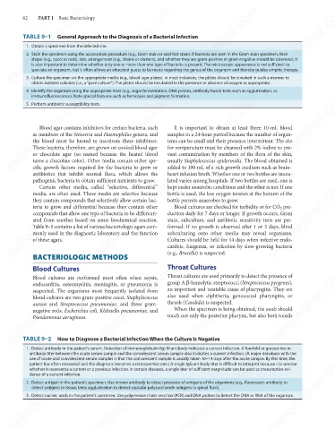
obtain isolated colonies (i.e., a “pure culture”). The plates should be incubated in the presence or absence of oxygen as appropriate.mebooksfree.com
mebooksfree.com
mebooksfree.com
mebooksfree.com
mebooksfree.com mebooksfree.com mebooksfree.com mebooksfree.com mebooksfree.com mebooksfree.com
62
PART I Basic Bacteriology
TABLE 9–1 General Approach to the Diagnosis of a Bacterial Infection
1. Obtain a specimen from the infected site.
2. Stain the specimen using the appropriate procedure (e.g., Gram stain or acid-fast stain). If bacteria are seen in the Gram stain specimen, their
shape (e.g., cocci or rods), size, arrangement (e.g., chains or clusters), and whether they are gram-positive or gram-negative should be observed. It
is also important to determine whether only one or more than one type of bacteria is present. The microscopic appearance is not sufficient to
speciate an organism, but it often allows an educated guess to be made regarding the genus of the organism and thereby guides empiric therapy.
3. Culture the specimen on the appropriate media (e.g., blood agar plates). In most instances, the plates should be streaked in such a manner to
4. Identify the organism using the appropriate tests (e.g., sugar fermentation, DNA probes, antibody-based tests such as agglutination, or
mebooksfree.com
mebooksfree.com mebooksfree.com mebooksfree.com samples in a 24-hour period because the number of organ- mebooksfree.com
mebooksfree.com
immunofluorescence). Note special features such as hemolysis and pigment formation.
5. Perform antibiotic susceptibility tests.
It is important to obtain at least three 10-mL blood
Blood agar contains inhibitors for certain bacteria, such
as members of the Neisseria and Haemophilus genera, and
isms can be small and their presence intermittent. The site
the blood must be heated to inactivate these inhibitors.
These bacteria, therefore, are grown on cooked blood agar
for venipuncture must be cleansed with 2% iodine to pre-
or chocolate agar (so named because the heated blood
usually Staphylococcus epidermidis. The blood obtained is
turns a chocolate color). Other media contain either spe-
cific growth factors required for the bacteria to grow or
added to 100 mL of a rich growth medium such as brain–
antibiotics that inhibit normal flora, which allows the vent contamination by members of the flora of the skin,
heart infusion broth. Whether one or two bottles are inocu-
lated varies among hospitals. If two bottles are used, one is
pathogenic bacteria to obtain sufficient nutrients to grow.
mebooksfree.com mebooksfree.com mebooksfree.com bottle permits anaerobes to grow. mebooksfree.com mebooksfree.com
mebooksfree.com
kept under anaerobic conditions and the other is not. If one
Certain other media, called “selective, differential”
media, are often used. These media are selective because
bottle is used, the low oxygen tension at the bottom of the
they contain compounds that selectively allow certain bac-
teria to grow and differential because they contain other
Blood cultures are checked for turbidity or for CO pro-
2
duction daily for 7 days or longer. If growth occurs, Gram
compounds that allow one type of bacteria to be differenti-
ated from another based on some biochemical reaction.
stain, subculture, and antibiotic sensitivity tests are per-
Table 9–3 contains a list of various bacteriologic agars com-
formed. If no growth is observed after 1 or 2 days, blind
subculturing onto other media may reveal organisms.
monly used in the diagnostic laboratory and the function
of these agars.
Cultures should be held for 14 days when infective endo-
carditis, fungemia, or infection by slow-growing bacteria
(e.g., Brucella) is suspected.
BACTERIOLOGIC METHODS
Blood Cultures
mebooksfree.com
mebooksfree.com mebooksfree.com mebooksfree.com Throat Cultures mebooksfree.com mebooksfree.com
Throat cultures are used primarily to detect the presence of
Blood cultures are performed most often when sepsis,
group A β-hemolytic streptococci (Streptococcus pyogenes),
endocarditis, osteomyelitis, meningitis, or pneumonia is
an important and treatable cause of pharyngitis. They are
suspected. The organisms most frequently isolated from
also used when diphtheria, gonococcal pharyngitis, or
blood cultures are two gram-positive cocci, Staphylococcus
thrush (Candida) is suspected.
aureus and Streptococcus pneumoniae, and three gram-
When the specimen is being obtained, the swab should
negative rods, Escherichia coli, Klebsiella pneumoniae, and
touch not only the posterior pharynx, but also both tonsils
Pseudomonas aeruginosa.
TABLE 9–2 How to Diagnose a Bacterial Infection When the Culture Is Negative
1. Detect antibody in the patient’s serum. Detection of immunoglobulin (Ig) M antibody indicates a current infection. A fourfold or greater rise in
antibody titer between the acute serum sample and the convalescent serum sample also indicates a current infection. (A major drawback with the
mebooksfree.com mebooksfree.com mebooksfree.com mebooksfree.com mebooksfree.com mebooksfree.com
use of acute and convalescent serum samples is that the convalescent sample is usually taken 10–14 days after the acute sample. By this time, the
patient has often recovered and the diagnosis becomes a retrospective one.) A single IgG antibody titer is difficult to interpret because it is unclear
whether it represents a current or a previous infection. In certain diseases, a single titer of sufficient magnitude can be used as presumptive evi-
dence of a current infection.
2. Detect antigen in the patient’s specimen. Use known antibody to detect presence of antigens of the organisms (e.g., fluorescent antibody to
detect antigens in tissue, latex agglutination to detect capsular polysaccharide antigens in spinal fluid).
3. Detect nucleic acids in the patient’s specimen. Use polymerase chain reaction (PCR) and DNA probes to detect the DNA or RNA of the organism.
mebooksfree.com mebooksfree.com mebooksfree.com mebooksfree.com mebooksfree.com mebooksfree.com