
Page 70 - TI Journal 18-1
P. 70
64 HUFFMAN ET. AL
mechanisms is provided below, followed by dis- Earlier studies initially identified this perivascular
cussion of advanced technological approaches to space where tracers move along periarterial spaces
enhance these models. (33,63,64,71,83). More recently, Iliff and colleagues
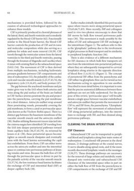
CSF is primarily produced in choroid plexuses of used in vivo two-photon microscopy to show that
the lateral, third, and fourth ventricles and is modestly ISF moves by bulk flow toward perivenous path-
regulated by the blood-CSF barrier (62). Functionally ways (36). This movement is made possible by the
similar to the blood brain barrier, the blood-CSF influx of CSF and subsequent CSF-ISF exchange in
barrier controls the production of CSF and its ionic the interstitium (Figure 1). The authors refer to this
and molecular composition while also serving as a as the ‘glymphatic’ pathway due to the involvement
pathway for solute and waste removal (18,39). CSF of glial processes in fluid transport and its similarity
travels through the ventricular system from the lateral to peripheral lymphatic function (36).
ventricles to the third and fourth ventricles, passing There is also evidence of a different mechanism
through the foramen of Magendie and Luschka where for ISF clearance in which bulk flow transports sol-
it mixes with existing fluid in the subarachnoid space utes from the interstitium into periarterial pathways,
(SAS) (54). The movement of CSF is then derived where they are subsequently transported to cervical
from multiple mechanisms, including hydrostatic lymphatics along arteries in the opposite direction
pressure gradients between CSF compartments and of blood flow (1,4,10,11) (Figure 1). The concept
sites of reabsorption (52), the pulsatility of the cardiac of periarterial ISF efflux from the parenchyma and
cycle and vascular smooth muscle (2,23,37,43,74), the CSF influx via glymphatic flow can be viewed as two
respiratory cycle (13,16,43), and body posture (47). mechanisms acting in opposition to one another.
The gap between the arachnoid membrane and pia Described in more detail below, it is important to note
mater gives way to the SAS where both arteries and that the precise anatomical differences between these
veins along the pial surface of the brain are bathed pathways are not yet fully understood. For the pur-
in CSF. Surface arteries penetrate the pia and project pose of this review, ‘perivascular space’ will broadly
into the parenchyma, carrying the pial membrane denote a single space between vascular endothelium
for a short distance. Astrocyte endfeet wrap around and astrocyte endfeet that permits the movement of
these penetrating vessels, presumably covering the CSF to, and ISF from, the parenchyma. ‘Glymphatic
pial membrane, to form a canal or perivascular space flow’ will represent the movement of subarachnoid
(also known as the Virchow-Robin space). There is a CSF along periarterial pathways, into the intersti-
distinct gap between the basement membrane of the tium to exchange with ISF, and then drained along
vascular smooth muscle and the astrocyte endfeet perivenous pathways.
(38). Studies utilizing fluorescent tracers injected into
the cisterna magna have identified CSF traveling from CLEARING THE BRAIN INTERSTITIUM
the SAS into deeper periarterial pathways toward
brain capillary beds (36,37,45,79). As reviewed by CSF Clearance
Jessen et al. (38), these periarterial spaces become Subarachnoid CSF can be transported to periph-
tighter as smooth muscle dissipates and eventually eral blood and lymphatics along four main routes of
joins with the basal lamina surrounding the capil- reabsorption: 1) arachnoid villi present in the dural
lary endothelium. From there, CSF can either move sinuses, 2) drainage pathways at the cranial nerves,
across the astrocyte endfeet and into the interstitial 3) nerve sheaths along spinal roots, and 4) the more
space or continue into perivenous pathways of drain- recently discovered lymphatic vessels in the meninges
ing venules and veins. Because the bulk flow of CSF (5,50,57). These drainage pathways are necessary
through the perivascular space is driven in part by for the removal of large solutes and metabolic waste
the pulsatile activity of the vascular smooth muscle dumped into ventricular and subarachnoid CSF.
(23,37,74), the low resistance basal lamina facilitates Clearance of the interstitial space relies in part on
the exchange of CSF and interstitial fluid (ISF) at either the bulk flow of ISF from the parenchyma to
deeper capillary beds (38). CSF compartments or the exchange of CSF and ISF