
Page 75 - TI Journal 18-1
P. 75
IMAGING THE PERIVASCULAR SPACE 69
AD (Figure 2). Furthermore, as animal studies have rhea (3,6). A recent clinical case study conducted by
successfully utilized contrast-enhanced MRI to exam- Eide and Ringstad also examined the distribution of
ine global fluid motion in rodents, these MR-based intrathecal injected gadolinium-based contrast agent
imaging techniques may have an important clinical throughout the brain, which the authors attribute to
application in the future. Current clinical practice glymphatic transport (17). As these results have yet
utilizes intrathecal injections of gadolinium-based to be further substantiated (17), conclusions cannot
contrasts to examine CSF leakage in disorders such as be fully drawn regarding future application. With
intracranial hypotension syndrome and CSF rhinor- that said, using gadolinium-based contrast materials
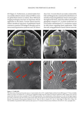
Figure 2. 7T MRI DPS
The observation of brain perivascular spaces. A representative slice of T2- weighted high resolution brain MR image at 7 T from a healthy
subject (a) and an AD patient (b). (c) and (d) are magnified images from the regions of interest marked by yellow squares in(a) and (b)
respectively. Red arrows show examples of PVSs with hyperintensity. (For interpretation of the references to color in this figure legend,
the reader is referred to the web version of the article: http://dx.doi.org/10.1016/j.jneumeth.2015.09.001). Reprinted by permission from
Elsevier: The Journal of Neuroscience Methods (Cai, K.; Tain, R.; Das, S.; Damen, F.C.; Sui, Y.; Valyi-Nagy, T.; Elliot, M.A.; Zhou, X.J.
The feasibility of quantitative MRI of perivascular spaces at 7T. Journal of Neuroscience Methods. 2015;256:151-156), copyright 2015.