
Page 20 - demo
P. 20
RESEARCH
LASER-IMAGING TECHNOLOGY
BROUGHT INTO FOCUS
Caltech’s Lihong Wang unveils improved method for peering inside living creatures
Caltech engineers have improved a technique for taking three-dimensional (3-D)
microscopic images of tissue, allowing them to see inside living creatures with
greater precision than before.
by Emily Velasco
he technology, called 3-D photoacoustic microscopy
T(PAM), bombards tissue with a laser beam. As the
energy in the laser light is absorbed, it causes the tissue
to vibra`te ultrasonically. Those vibrations are picked up
by sensors and used to assemble an image of the tissue’s
internal structures in a process similar to ultrasound
imaging.
The technique was invented by Lihong Wang, Caltech’s
Professor of Medical Engineering and Electrical Engineering,
and his team in the Caltech Optical Imaging Laboratory,
part of the Andrew and Peggy Cherng Department of
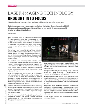
Medical Engineering in the Division of Engineering and Jiamiao Yang, a postdoctoral scholar in the Caltech Optical Imaging
Applied Science. Laboratory, adjusts a photoacoustic microscope.
One constraint of the technology to this point has been Credit: Caltech
its limited depth of field—the range at which objects are These modifications give SIR-PAM a depth of field 32 times
in focus. This phenomenon would be familiar to anyone who larger than what PAM could achieve while also improving its
has used a camera. When the camera is focused on a nearby resolution to as small as 90 nanometers (1/1000th the width
object, objects in the background will be blurry. When the of a human hair).
camera is focused on something in the distance, nearby
objects are blurry. “This gives us the ability to look through opaque materials
and see what’s inside,” Wang says. “It’s like an extension of
While such blurring can add an artsy flair on Instagram, the human eye, like Superman’s X-ray vision.”
it is not desirable in 3-D medical imaging, so Wang and
his team set out to tweak their technology to minimize “Photoacoustics is unique,” he says. “It can be scaled to image
the effect. In a paper published in the October 3 issue of everything from structures inside a cell all the way up to an
Nature Communications, they describe a modified form of entire organism, affording an unprecedented opportunity
the technology they’re calling spatially invariant resolution for omniscale biological research with consistent imaging
photoacoustic microscopy, or SIR-PAM. contrast.”
20 SIR-PAM builds on previous PAM technology by pre-processing The paper is titled “Motionless volumetric photoacoustic
the laser beam with a specialized optical chip found in certain microscopy with spatially invariant resolution.”Wang’s other
types of TVs and projectors. The chip splits the beam in two, co-authors are Caltech researchers Jiamiao Yang, Yuecheng
and each of those beams bombards the object to be imaged Shen, and Pengfei Hai, Lei Gong, Xiao Xu, and Yuta Suzuki,
from a different angle. researchers from Wang’s former lab at Washington University
in St. Louis.
When the beams cross inside the object, they create precise
interference patterns that provide acoustic signatures Funding for the research was provided by the National
needed to construct a clear 3-D image of internal structures Institutes of Health.
throughout the scanned area.
www.ghmediabusiness.com
| Global MDA Journal | NOV-DEC 2017