
Page 244 - Ultimate Visual Dictionary (DK)
P. 244
THE HUMAN BODY
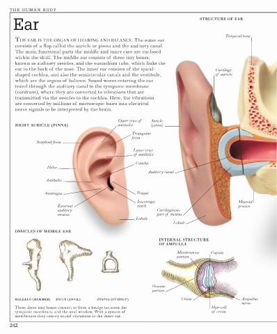
Ear STRUCTURE OF EAR
Temporal bone
THE EAR IS THE ORGAN OF HEARING AND BALANCE. The outer ear
consists of a flap called the auricle or pinna and the auditory canal.
The main functional parts-the middle and inner ears-are enclosed
within the skull. The middle ear consists of three tiny bones,
known as auditory ossicles, and the eustachian tube, which links the
ear to the back of the nose. The inner ear consists of the spiral- Cartilage
shaped cochlea, and also the semicircular canals and the vestibule, of auricle
which are the organs of balance. Sound waves entering the ear
travel through the auditory canal to the tympanic membrane
(eardrum), where they are converted to vibrations that are
transmitted via the ossicles to the cochlea. Here, the vibrations
are converted by millions of microscopic hairs into electrical
nerve signals to be interpreted by the brain.
Upper crux of Auricle
RIGHT AURICLE (PINNA) antihelix (pinna)
Triangular
fossa
Scaphoid fossa
Lower crux
of antihelix
Concha
Helix
Auditory canal
Antihelix
Antitragus Tragus
Intertragic Mastoid
External notch process
auditory Cartilaginous
meatus part of meatus
Lobule
Lobule
OSSICLES OF MIDDLE EAR
INTERNAL STRUCTURE
OF AMPULLA
Membranous Cupula
portion
Osseous
portion
MALLEUS (HAMMER) INCUS (ANVIL) STAPES (STIRRUP) Crista Ampullar
nerve
These three tiny bones connect to form a bridge between the Hair cell
tympanic membrane and the oval window. With a system of of crista
membranes they convey sound vibrations to the inner ear.
242