
Page 521 - First Aid for the USMLE Step 1 2020, Thirtieth edition [MedicalBooksVN.com]_Neat
P. 521
Musculoskeletal, skin, and connective tissue ` dERmatology Musculoskeletal, skin, and connective tissue ` dERmatology section iii 477
Common skin disorders
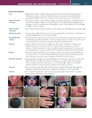
Acne Multifactorial etiology— sebum/androgen production, abnormal keratinocyte desquamation,
Cutibacterium acnes colonization of the pilosebaceous unit (comedones), and inflammation
(papules/pustules A, nodules, cysts). Treatment: retinoids, benzoyl peroxide, and antibiotics.
Atopic dermatitis Type I hypersensitivity reaction. Pruritic eruption, commonly on skin flexures. Associated with other
(eczema) atopic diseases (asthma, allergic rhinitis, food allergies); serum IgE. Mutations in filaggrin gene
predispose (via skin barrier dysfunction). Often appears on face in infancy B and then in antecubital
fossa C in children and adults.
Allergic contact Type IV hypersensitivity reaction secondary to contact allergen (eg, nickel D, poison ivy, neomycin E ).
dermatitis
Melanocytic nevus Common mole. Benign, but melanoma can arise in congenital or atypical moles. Intradermal nevi
are papular F . Junctional nevi are flat macules G.
Pseudofolliculitis Foreign body inflammatory facial skin disorder characterized by firm, hyperpigmented papules and
barbae pustules that are painful and pruritic. Located on cheeks, jawline, and neck. Commonly occurs as
a result of shaving (“razor bumps”), primarily affects African-American males.
Psoriasis Papules and plaques with silvery scaling H, especially on knees and elbows. Acanthosis with
parakeratotic scaling (nuclei still in stratum corneum), Munro microabscesses. stratum
spinosum, stratum granulosum. Auspitz sign ( I )—pinpoint bleeding spots from exposure of
dermal papillae when scales are scraped off. Associated with nail pitting and psoriatic arthritis.
Rosacea Inflammatory facial skin disorder characterized by erythematous papules and pustules J , but no
comedones. May be associated with facial flushing in response to external stimuli (eg, alcohol,
heat). Phymatous rosacea can cause rhinophyma (bulbous deformation of nose).
Seborrheic keratosis Flat, greasy, pigmented squamous epithelial proliferation of immature keratinocytes with keratin-
filled cysts (horn cysts) K . Looks “stuck on.” Lesions occur on head, trunk, and extremities.
Common benign neoplasm of older persons. Leser-Trélat sign L —rapid onset of multiple
seborrheic keratoses, indicates possible malignancy (eg, GI adenocarcinoma).
Verrucae Warts; caused by low-risk HPV strains. Soft, tan-colored, cauliflower-like papules M. Epidermal
hyperplasia, hyperkeratosis, koilocytosis. Condyloma acuminatum on anus or genitals N.
Urticaria Hives. Pruritic wheals that form after mast cell degranulation O. Characterized by superficial
dermal edema and lymphatic channel dilation.
A B C D E
F G H I J
K L M N O
FAS1_2019_11-Musculo.indd 477 11/7/19 5:24 PM