
Page 112 - fbkCardioDiabetes_2017
P. 112
88 Peripheral Vascular Disease
activity such as walking and disappears after several Classification of PVD
minutes of rest
Grade Category Clinical Discription
0 0 Asymptomatic
i 1 Mild claudication
ii 2 Mod-Severe claudication
iii 3 Sev claudication
iv 4 Ulceration or gangrene
Fig.4. The Rose angina and claudication questionnaire and
the Edinburgh claudication questionnaire can be used to as-
sist clinicians with screening patients for PAD 2&3 .
Physical examination includes measurement of blood
pressure in both arms, cardiac auscultation for heart
rate and rhythm, auscultation for carotid, subclavi-
an, abdominal and femoral artery bruits, abdominal
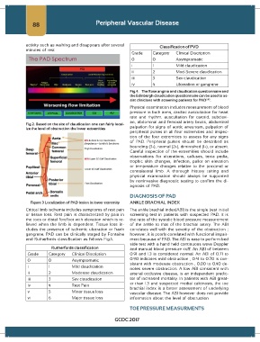
Fig.2. Based on the site of claudication one can fairly local-
ize the level of obstruction the lower extremities palpation for signs of aortic aneurysm, palpation of
peripheral pulses in all four extremities and inspec-
tion of the four extremities to assess for any signs
of PAD. Peripheral pulses should be described as
bounding (3+), normal (2+), diminished (1+), or absent.
Careful inspection of the extremities should include
observations for ulcerations, calluses, tenia pedia,
trophic skin changes, infection, pallor on elevation
or temperature changes relative to the proximal or
contralateral limb. A thorough history taking and
physical examination should always be supported
by noninvasive diagnostic testing to confirm the di-
agnosis of PAD.
DIAGNOSIS OF PAD
Figure 3 Localization of PAD lesion in lower extremity ANKLE BRACHIAL INDEX
Critical limb ischemia includes symptoms of rest pain The ankle brachial index(ABI) is the single best initial
or tissue loss. Rest pain is characterized by pain in screening test in patients with suspected PAD. It is
the toes or distal forefoot with elevation which is re- the ratio of the systolic blood pressure measurement
lieved when the limb is dependent. Tissue loss in- of the ankle to that of the brachial artery. The ABI
cludes the presence of ischemic ulceration or frank correlates well with the severity of the obstruction ;
gangrene. PAD can be clinically staged by Fontaine however, it is poorly correlated with functional impair-
and Rutherfords classification as follows Fig.4. ment because of PAD. The ABI is easy to perform bed
side test with a hand held continuous wave Doppler
Rutherfords classification and manual blood pressure cuff. An ABI of between
Grade Category Clinical Discription 0.91 and 1.3 is considered normal. An ABI of 0.71 to
0 0 Asymptomatic 0.90 indicates mild obstruction , 0.41 to 0.70 is con-
sistent with moderate obstruction , 0.00 to 0.40 de-
i 1 Mild claudication
notes severe obstruction. A low ABI consistent with
ii 2 Moderate claudication arterial occlusive disease, is an independent predic-
iii 3 Sev claudication tor of increased mortality. In patients with ABI great-
er than 1.3 and suspected medial calcinosis, the toe
iv 4 Rest Pain
brachial index is a better assessment of underlying
v 5 Minor tissue loss vascular disease. The ABI however does not provide
vi 6 Major tissue loss information about the level of obstruction
TOE PRESSURE MEASURMENTS
GCDC 2017