
Page 383 - fbkCardioDiabetes_2017
P. 383
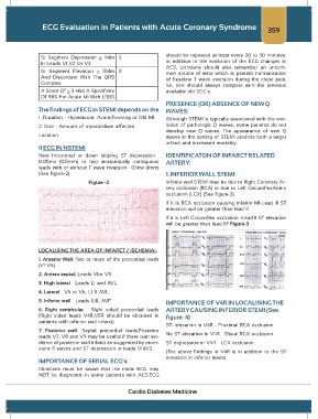
ECG Evaluation in Patients with Acute Coronary Syndrome                                   359


                                                                    should be repeated at least every 20 to 30 minutes.
                 St Segment  Depression  > 1Mm  3
                 In Leads V1,V2 Or V3                               In addition to the evolution of the  ECG  changes in
                                                                    ACS, clinicians  should also  remember  an uncom-
                 St  Segment Elevation  >  5Mm  2                   mon source of error  which  is  pseudo  normalisation
                 And Discordant  With  The QRS                      of baseline  T wave inversion  during the chest pain.
                 Complex                                            So, one should always compare  with  the  previous
                 A Score Of > 3 Had A Specificity                   available old ECG’s.
                 Of 98% For Acute MI With LBBB
                                                                    PRESENCE (OR) ABSENCE OF NEW Q
                 The findings of ECG in STEMI depends on the        WAVES:
                 1. Duration - Hyperacute, Acute,Evolving or Old MI  Although STEMI is typically associated with the evo-

                 2. Size - Amount of myocardium affected            lution of pathologic Q waves, some patients do not
                                                                    develop  new Q waves.  The  appearance  of  new Q
                 Location                                           waves in the setting of STEMI predicts both a larger
                                                                    infarct and increased mortality².
                 II ECG IN NSTEMI
                 New  horizontal  or  down  sloping  ST  depression  ≥  IDENTIFICATON OF INFARCT RELATED
                 0.05mv  (0.5mm) in two anatomically  contiguous    ARTERY:
                 leads with or without T wave inversion ≥ 0.1mv (1mm)
                 (See figure-2)                                     1. INFERIOR WALL STEMI:

                                     Figure -2                      Inferior wall STEMI may be due to Right Coronary Ar-
                                                                    tery occlusion (RCA) or due to Left CircumflexArtery
                                                                    occlusion (LCX) (See figure-3)
                                                                    If it is RCA occlusion causing inferior MI-Lead III ST
                                                                    elevation will be greater than lead II³

                                                                    If it is Left Circumflex occlusion -Lead II ST elevation
                                                                    will be greater than lead III Figure-3
                                                                                            4


                 LOCALISING THE AREA OF INFARCT / ISCHEMIA:-
                 1. Anterior Wall: Two or  more of the precordial  leads
                 (V1-V6)

                 2. Antero septal: Leads V1to V3
                 3. High lateral – Leads LI and AVL.
                 4. Lateral – V4 to V6, LI & AVL.
                 5. Inferior wall – Leads II,III, AVF               IMPORTANCE OF V4R IN LOCALISING THE
                 6. Right  ventricular  – Right sided  precordial  leads  ARTERY CAUSING INFERIOR STEMI (See
                 (Right  sided leads V4R,V5R  should be obtained  in   figure- 4)
                 patients with inferior wall infarct)
                                                                    ST- elevation in V4R - Proximal RCA occlusion
                 7. Posterior  wall- Septal precordial  leads.Posterior
                 leads V7, V8 and V9 may be useful if there isan evi-  No ST elevation in V4R – Distal RCA occlusion.
                 dence of posterior wall infarct as suggested by prom-  ST depression in V4R – LCX occlusion.
                 inent R waves and ST depression in leads V1&V2.
                                                                    (The above  findings  in V4R is  in addition to the ST
                                                                    elevation in inferior leads)
                 IMPORTANCE OF SERIAL ECG’s:
                 Clinicians must be  aware  that  the initial ECG may
                 NOT  be diagnostic in some patients with  ACS.ECG


                                                    Cardio Diabetes Medicine