
Page 387 - fbkCardioDiabetes_2017
P. 387
Strain and Strain Rate Imaging in Early Detection of Ventricular 363
Systolic Dysfunction - is This The Best Investigation?
New Methods For Evaluation of LV Systolic Decrease of the dimension (shortening of the wall in
Function longitudinal direction during systole, or decrease of
the circumferential dimension during systole, as well
Newer echocardiographic techniques, such as TDI as thinning of the wall during diastole) is marked with
and deformation imaging gives better understand- a negative number ( negative sign –) and increase
ing and evaluation of the complex mechanism of of the dimension (lengthening of the wall in a lon-
cardiac contraction and relaxation. Evaluation of LV gitudinal direction during diastole, or increase of the
longitudinal systolic dynamics has become crucial in circumferential dimension during systole, as well as
the assessment of LV systolic function and superi- thickening of the wall during systole) is marked with
or value in comparison with traditional measures [4] a positive number ( positive sign +).
and is evaluated using Tissue Doppler and speckle
tracking techniques utilizing strain (S) and strain rate
(SR) imaging thus giving a more detailed information Types of Strains
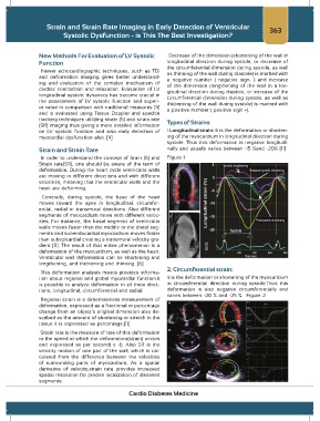
on LV systolic function and also early detection of 1.Longitudinal strain: It is the deformation or shorten-
myocardial dysfunction also. [4] ing of the myocardium in longitudinal direction during
systole .Thus this deformation is negative longitudi-
Strain and Strain Rate nally and usually varies between -15 %and -20% [11]
In order to understand the concept of Srain (S) and Figure-1
Strain rate(SR), one should be aware of the term of
deformation. During the heart cycle ventricular walls
are moving in different directions and with different
velocities, meaning that the ventricular walls and the
heart are deforming.
Generally, during systole, the base of the heart
moves toward the apex in longitudinal, circumfer-
ential, radial or transmural directions. Also different
segments of myocardium move with different veloc-
ities. For instance, the basal segment of ventricular
walls moves faster than the middle or the distal seg-
ments and subendocardial myocardium moves faster
than subepicardial creating a transmural velocity gra-
dient [5]. The result of that entire phenomenon is a
deformation of the myocardium, as well as the heart.
Ventricular wall deformation can be shortening and
lengthening, and thickening and thinning. [6]
2. Circumferential strain:
This deformation analysis mainly provides informa-
tion about regional and global myocardial function.It It is the deformation or shortening of the myocardium
is possible to analyze deformation in all three direc- in circumferential direction during systole.Thus this
tions, longitudinal, circumferential and radial. deformation is also negative circumferentially and
varies between -20 % and -25 %. Figure 2
Regional strain is a dimensionless measurement of
deformation, expressed as a fractional or percentage
change from an object’s original dimension also de-
scribed as the amount of shortening or stretch in the
tissue It is expressed as percentage.[11]
Strain rate is the measure of rate of this deformation
or the speed at which the deformation(strain) occurs
and expressed as per second( s -1). Also SR is the
velocity motion of one part of the wall, which is cal-
culated from the difference between the velocities
of surrounding parts of myocardium, As a spatial
derivative of velocity,strain rate provides increased
spatial resolution for precise localization of diseased
segments.
Cardio Diabetes Medicine