
Page 291 - Critical Care Nursing Demystified
P. 291
276 CRITICAL CARE NURSING DeMYSTIFIED
therapy is initiated first and blood and colloids are considered if the response
of the heart rate, blood pressure, and baseline laboratory values do not improve.
Blood replacement is usually determined by changes in the patient’s hemoglo-
bin and hematocrit levels. Although resources vary, when the patient’s hemo-
globin drops to 8 g/dL and the patient has other associated symptoms like
unstable hemodynamic parameters, blood should be administered. Typically,
packed red blood cells or whole blood is given in the traumatized patient, with
whole blood being reserved for patients with coagulation problems like acute
gastrointestinal bleeding from esophageal varices.
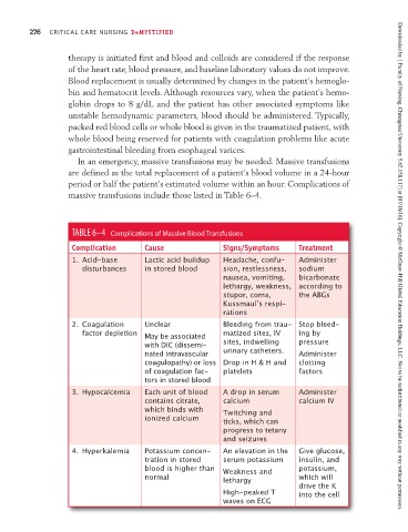
In an emergency, massive transfusions may be needed. Massive transfusions
are defined as the total replacement of a patient’s blood volume in a 24-hour
period or half the patient’s estimated volume within an hour. Complications of
massive transfusions include those listed in Table 6–4.
TABLE 6–4 Complications of Massive Blood Transfusions
Complication Cause Signs/Symptoms Treatment
1. Acid-base Lactic acid buildup Headache, confu- Administer
disturbances in stored blood sion, restlessness, sodium Downloaded by [ Faculty of Nursing, Chiangmai University 5.62.158.117] at [07/18/16]. Copyright © McGraw-Hill Global Education Holdings, LLC. Not to be redistributed or modified in any way without permission.
nausea, vomiting, bicarbonate
lethargy, weakness, according to
stupor, coma, the ABGs
Kussmaulʼs respi-
rations
2. Coagulation Unclear Bleeding from trau- Stop bleed-
factor depletion May be associated matized sites, IV ing by
with DIC (dissemi- sites, indwelling pressure
nated intravascular urinary catheters. Administer
coagulopathy) or loss Drop in H & H and clotting
of coagulation fac- platelets factors
tors in stored blood
3. Hypocalcemia Each unit of blood A drop in serum Administer
contains citrate, calcium calcium IV
which binds with Twitching and
ionized calcium ticks, which can
progress to tetany
and seizures
4. Hyperkalemia Potassium concen- An elevation in the Give glucose,
tration in stored serum potassium insulin, and
blood is higher than Weakness and potassium,
normal lethargy which will
drive the K
High-peaked T into the cell
waves on ECG