
Page 125 - Color_Atlas_of_Physiology_5th_Ed._-_A._Despopoulos_2003
P. 125
.
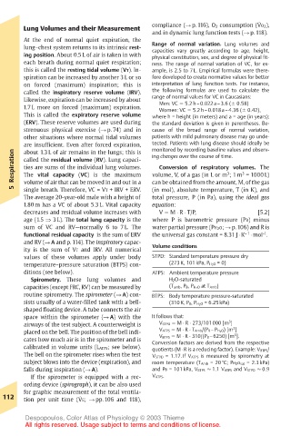
Lung Volumes and their Measurement compliance (! p. 116), O 2 consumption (VO 2),
and in dynamic lung function tests (! p. 118).
At the end of normal quiet expiration, the
lung–chest system returns to its intrinsic rest- Range of normal variation. Lung volumes and
capacities vary greatly according to age, height,
ing position. About 0.5 L of air is taken in with physical constitution, sex, and degree of physical fit-
each breath during normal quiet respiration; ness. The range of normal variation of VC, for ex-
this is called the resting tidal volume (VT). In- ample, is 2.5 to 7 L. Empirical formulas were there-
spiration can be increased by another 3 L or so fore developed to create normative values for better
on forced (maximum) inspiration; this is interpretation of lung function tests. For instance,
called the inspiratory reserve volume (IRV). the following formulas are used to calculate the
Likewise, expiration can be increased by about range of normal values for VC in Caucasians:
Men: VC ! 5.2 h–0.022a–3.6 (" 0.58)
1.7 L more on forced (maximum) expiration. Women: VC ! 5.2 h–0.018a–4.36 (" 0.42),
This is called the expiratory reserve volume where h = height (in meters) and a = age (in years);
(ERV). These reserve volumes are used during the standard deviation is given in parentheses. Be-
strenuous physical exercise (! p. 74) and in cause of the broad range of normal variation,
other situations where normal tidal volumes patients with mild pulmonary disease may go unde-
are insufficient. Even after forced expiration, tected. Patients with lung disease should ideally be
monitored by recording baseline values and observ-
Respiration called the residual volume (RV). Lung capaci- ing changes over the course of time. 3
about 1.3 L of air remains in the lungs; this is
ties are sums of the individual lung volumes.
Conversion of respiratory volumes. The
3
The vital capacity (VC) is the maximum
volume, V, of a gas (in L or m ; 1 m = 1000 L)
volume of air that can be moved in and out in a
5 single breath. Therefore, VC = VT + IRV + ERV. can be obtained from the amount, M, of the gas
(in mol), absolute temperature, T (in K), and
The average 20-year-old male with a height of total pressure, P (in Pa), using the ideal gas
1.80 m has a VC of about 5.3 L. Vital capacity equation:
decreases and residual volume increases with V ! M " R " T/P, [5.2]
age (1.5 ! 3 L). The total lung capacity is the where P is barometric pressure (PB) minus
sum of VC and RV—normally 6 to 7 L. The water partial pressure (PH 2 O; ! p. 106) and R is
-1
functional residual capacity is the sum of ERV the universal gas constant = 8.31 J " K – 1 " mol .
and RV (! A and p. 114). The inspiratory capac- Volume conditions
ity is the sum of VT and IRV. All numerical
values of these volumes apply under body STPD: Standard temperature pressure dry
temperature–pressure saturation (BTPS) con- (273 K, 101 kPa, P H 2 O = 0)
ditions (see below). ATPS: Ambient temperature pressure
Spirometry. These lung volumes and H 2O-saturated
capacities (except FRC, RV) can be measured by (T amb, P B, P H 2 O at T Amb)
routine spirometry. The spirometer (! A) con- BTPS: Body temperature pressure-saturated
sists usually of a water-filled tank with a bell- (310 K, P B, P H 2 O = 6.25 kPa)
shaped floating device. A tube connects the air
space within the spirometer (! A) with the It follows that:
3
airways of the test subject. A counterweight is V STPD ! M " R " 273/101000 [m ] 3
placed on the bell. The position of the bell indi- V ATPS ! M " R " T Amb/(P B –P H 2 O) [m ] 3
V BTPS ! M " R " 310/(P B –6250) [m ].
cates how much air is in the spirometer and is Conversion factors are derived from the respective
calibrated in volume units (L ATPS; see below). quotients (M " R is a reducing factor). Example: V BTPS/
The bell on the spirometer rises when the test V STPD = 1.17. If V ATPS is measured by spirometry at
subject blows into the device (expiration), and room temperature (T Amb = 20 #C; PH 2 O sat = 2.3 kPa)
falls during inspiration (! A). and PB = 101 kPa, V BTPS ! 1.1 V ATPS and V STPD ! 0.9
If the spirometer is equipped with a rec- V ATPS.
ording device (spirograph), it can be also used
for graphic measurement of the total ventila-
.
112 tion per unit time (VE; ! pp. 106 and 118),
Despopoulos, Color Atlas of Physiology © 2003 Thieme
All rights reserved. Usage subject to terms and conditions of license.