
Page 203 - Color_Atlas_of_Physiology_5th_Ed._-_A._Despopoulos_2003
P. 203
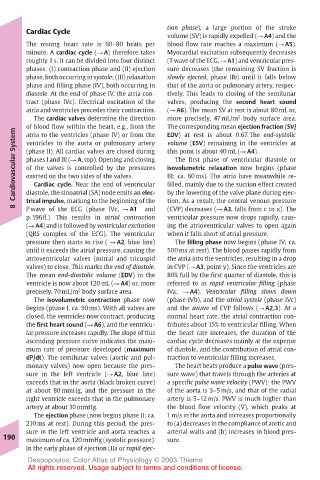
Cardiac Cycle                   tion phase), a large portion of the stroke
                                       volume (SV) is rapidly expelled (! A4) and the
       The resting heart rate is 60–80 beats per  blood flow rate reaches a maximum (! A5).
       minute. A cardiac cycle (! A) therefore takes  Myocardial excitation subsequently decreases
       roughly 1 s. It can be divided into four distinct  (T wave of the ECG, ! A1) and ventricular pres-
       phases: (I) contraction phase and (II) ejection  sure decreases (the remaining SV fraction is
       phase, both occurring in systole; (III) relaxation  slowly ejected, phase IIb) until it falls below
       phase and filling phase (IV), both occurring in  that of the aorta or pulmonary artery, respec-
       diastole. At the end of phase IV, the atria con-  tively. This leads to closing of the semilunar
       tract (phase IVc). Electrical excitation of the  valves, producing the second heart sound
       atria and ventricles precedes their contraction.  (! A6). The mean SV at rest is about 80 mL or,
         The cardiac valves determine the direction  more precisely, 47 mL/m body surface area.
                                                       2
       of blood flow within the heart, e.g., from the  The corresponding mean ejection fraction (SV/
    Cardiovascular System  (phase II). All cardiac valves are closed during  this point is about 40 mL (! A4).
                                       EDV) at rest is about 0.67. The end-systolic
       atria to the ventricles (phase IV) or from the
                                       volume (ESV) remaining in the ventricles at
       ventricles to the aorta or pulmonary artery
       phases I and III (! A, top). Opening and closing
                                        The first phase of ventricular diastole or
       of the valves is controlled by the pressures
                                       isovolumetric relaxation now begins (phase
       exerted on the two sides of the valves.
                                       III; ca. 60 ms). The atria have meanwhile re-
         Cardiac cycle. Near the end of ventricular
                                       filled, mainly due to the suction effect created
                                       tion. As a result, the central venous pressure
       trical impulse, marking to the beginning of the
    8  diastole, the sinoatrial (SA) node emits an elec-  by the lowering of the valve plane during ejec-
                                       (CVP) decreases (! A3, falls from c to x). The
       P wave of the ECG (phase IVc, ! A1
                                  and
       p. 196ff.). This results in atrial contraction  ventricular pressure now drops rapidly, caus-
       (! A4) and is followed by ventricular excitation  ing the atrioventricular valves to open again
       (QRS complex of the ECG). The ventricular  when it falls short of atrial pressure.
       pressure then starts to rise ( ! A2, blue line)  The filling phase now begins (phase IV; ca.
       until it exceeds the atrial pressure, causing the  500 ms at rest). The blood passes rapidly from
       atrioventricular valves (mitral and tricuspid  the atria into the ventricles, resulting in a drop
       valves) to close. This marks the end of diastole.  in CVP (! A3, point y). Since the ventricles are
       The mean end-diastolic volume (EDV) in the  80% full by the first quarter of diastole, this is
       ventricle is now about 120 mL (! A4) or, more  referred to as rapid ventricular filling (phase
                   2
       precisely, 70 mL/m body surface area.  IVa; ! A4). Ventricular filling slows down
         The isovolumetric contraction phase now  (phase IVb), and the atrial systole (phase IVc)
       begins (phase I, ca. 50 ms). With all valves are  and the awave of CVP follows (! A2,3). At a
       closed, the ventricles now contract, producing  normal heart rate, the atrial contraction con-
       the first heart sound (! A6), and the ventricu-  tributes about 15% to ventricular filling. When
       lar pressure increases rapidly. The slope of this  the heart rate increases, the duration of the
       ascending pressure curve indicates the maxi-  cardiac cycle decreases mainly at the expense
       mum rate of pressure developed (maximum  of diastole, and the contribution of atrial con-
       dP/dt). The semilunar valves (aortic and pul-  traction to ventricular filling increases.
       monary valves) now open because the pres-  The heart beats produce a pulse wave (pres-
       sure in the left ventricle (! A2, blue line)  sure wave) that travels through the arteries at
       exceeds that in the aorta (black broken curve)  a specific pulse wave velocity (PWV): the PWV
       at about 80 mmHg, and the pressure in the  of the aorta is 3–5 m/s, and that of the radial
       right ventricle exceeds that in the pulmonary  artery is 5–12 m/s. PWV is much higher than
                                                       .
       artery at about 10 mmHg.        the blood flow velocity (V), which peaks at
         The ejection phase (now begins phase II; ca.  1 m/s in the aorta and increases proportionally
       210 ms at rest). During this period, the pres-  to (a) decreases in the compliance of aortic and
       sure in the left ventricle and aorta reaches a  arterial walls and (b) increases in blood pres-
  190  maximum of ca. 120 mmHg (systolic pressure).  sure.
       In the early phase of ejection (IIa or rapid ejec-
       Despopoulos, Color Atlas of Physiology © 2003 Thieme
       All rights reserved. Usage subject to terms and conditions of license.